

 PDF
PDF ePub
ePub Citation
 Print
Print


INTRODUCTION
Henri Francois Secretan first reported 11 cases of workmen who had a hard swelling of the dorsum of the hand after trauma in 1901 (1). Named after Secrtan, the disease usually occurred after minor trauma of the hand without osseous fracture or repetitive self-inflicted injury, without definitive etiology and pathophysiology. Peritendinous fibrosis on the extensor tendons of the hand is one of the widely recognized histologic feature. The earlier reports indicate the general involvement of adults over the age of 30 and with trauma or repetitive working histories (1-5). Secretan's disease, is not only limited to the hands also in the upper and lower extremities including feet was reported (6, 7).
Since, magnetic resonance imaging (MRI) analyses of Secretan's disease is sparsely studied in the literature. Here, we report a case of Secretan's disease with MRI findings.
CASE REPORT
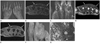
A 73-year-old woman presented with painful hard swelling on the dorsum of both hands. The patient complained an approximate 30-years history of intermittent, self-limiting swelling on both hands and since the past year, swelling and pain was gradually progressed. The symptoms were predominant on the left hand. She was a full-time home-maker with no history of recent hand trauma. She has also not suffered from any other medical illness including psychiatric problems. Investigations revealed hard swelling on the dorsum of both hands with mild erythematous skin change (Fig. 1A).
Laboratory investigations including common serum rheumatologic tests (e.g., erythrocyte sedimentation rate, C-reactive protein, rheumatologic factor and anti-nuclear antibody) were unremarkable. Radiographs of the hands showed no distinct abnormal finding except non-specific soft tissue swelling on the dorsum of the hands.
MRI was performed to further characterize the soft tissue lesions. T2-weighted fat suppression and T1-weighted images depicted an ill-defined, infiltrative mass like lesion with diffuse hypointensity in the subcutaneous layer from second to forth metacarpal area. The lesion extended into the peritendinous area of the extensor tendons at the level of the metacarpophalangeal joints. Interstitial edema and swelling around the lesion were also seen. On contrast-enhanced fat-suppressed T1-weighted images, there was no significant enhancement within the lesion (Fig. 1B-E).
The patient underwent excisional biopsy which revealed a yellowish, stony hard infiltrative lesion with very poorly defined margin in the subcutaneous layer and around the extensor tendon sheath, however, with no significant adhesion between the lesion and the tendons (Fig. 1F). The histologic sections for the lesion demonstrated a dense fibrotic lesion with the depositions of hyaline materials and prominent fatty degenerations. There was no evidence indicating a neoplastic pathology (Fig. 1G).
Despite though the patient reported no obvious trauma or self-injury history, considering the general causal relationship between 'light' trauma and this disease, it may be difficult to identify the relevance of trauma in this case. Therefore, we and our orthopedic surgery and surgical pathology teams concluded the diagnosis to of Secretan's disease based on clinical, radiologic and histologic findings.
DISCUSSION
Since its first report of Secretan's disease, the postulated three essential etiologies for this disease are 1) reactive peritendinous fibrosis induced by organizing hematoma after true light trauma, 2) swelling caused by hypersympathetic process related to an injury with neurovascular or neurolymphatic changes, and 3) repeated self-inflicted injury for secondary gain or as the product of an underlying psychiatric problem resulting in factitious edema or hyperplastic fibrosis (1). The disease also shows variable clinical manifestations from soft swelling as a benign course to a hyperplastic course with persistent and hard fibrosis that limits the range of motion of fingers (1).
Secretan's disease was diagnosed based on medical history and physical examination, however, further imaging assessments such as MRI and ultrasonography have not been generally adapted.
Only Whitney and Jones (2) described MRI findings of 3 cases of clinically suspected Secretan's disease. Common MRI findings of the cases were depicted as abnormal soft tissue infiltration surrounding the extensor tendons including radiological description of tendon swelling and focal scarring consistent with peritendinous fluid and ganglion tissue. The surgical specimens of two cases showed fibrous tissue with cystic and myxoid degeneration similar to ganglia formation. There seems to be slightly different from previously noted histological results focusing on the peritendinous fibrosis due to ganglia with chronic ruptured state may result in peritendinous fibrosis and clinical manifestations similar to Secretan's disease.
Major MRI findings of this case are well correlated with histologic results, representing the presence of prominent fibrosis within the lesion with little evidence of the ganglion or recently developed hematoma and we assume that the image findings could be quite variable depending on the course of the disease.
To the best of our knowledge, this disease generally occurs in the hand having been afflicted with a preceding traumatic injury. Interestingly, however, the patient in our case presented symmetrical hard swelling on both hands and had no definite trauma history. However, it may be difficult to prove the exact association of this case with trauma due to her long-term illness.
The differential diagnosis of Secretan's disease includes the conditions that can cause recurrent or chronic hand edema. Systemic illnesses such as kidney disease, congestive heart failure and liver diseases are excluded through the clinical assessment. In the musculoskeletal area, most arthropathies and tenosynovial pathologies involving the hands should be differentiated, which include various rheumatologic disorders from common rheumatoid arthritis to remitting seronegative symmetrical synovitis with pitting edema, a widely known rheumatologic pathology. Among the neoplastic conditions occurring around the tendinous structures in the adult population, giant cell tumor of tendon sheath or fibroma of tendon sheath should also be considered as a differential diagnosis. MRI may be helpful to characterize and discriminate this rare disease from others.
In conclusion, the present report presents a rare case of Secretan's disease with characteristic MRI findings including ill-defined infiltrative lesions in the dorsal subcutaneous layer of the hand with homogenous dark signal intensity on T1 and T2-weighted images and poor enhancement on postcontrast scans suggesting the fibrotic component within the lesion.
 XML Download