

 PDF
PDF ePub
ePub Citation
 Print
Print


INTRODUCTION
Metastatic pulmonary calcification (MPC) tends to be presented in primary or secondary hyperparathyroidism, such as chronic renal failure. CT scan is a good imaging modality for the detection of a small amount of pulmonary calcification, ground glass opacity (GGO), and nodular opacity in upper- and mid-lung fields. For the reason that findings of early stage of MPC are atypical and similar to those of other diseases, such as pulmonary edema and pneumonia, it is hard to detect and diagnose the MPC early (1, 2). Additional imaging modalities and transbronchial lung biopsy may be obvious means for detection of MPC (1, 3).
We report on an interesting case in which an initial CT scan showed abnormal findings and thus, following up on the CT scan for five years for typical findings of MPC combined with alveolar hemorrhage.
CASE REPORT
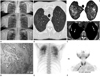
A 50-year-old male diagnosed with end-stage renal disease was admitted with consistent nausea and emesis. He had renal transplantation 14 years ago. After the transplantation, the patient developed chronic rejections and was treated with an immunosuppressive agent. Since then, he has been dialyzed for seven years. Five years ago, the patient was admitted to our hospital with fever and mild dyspnea. At that time, the laboratory findings indicated leukocytosis and hypercalcemia. Plain radiograph of the chest showed ill-defined nodular opacities in both upper lungs (Fig. 1A).
On this admission, laboratory findings indicated increasing BUN/Cr level of 101.3 (8-20) and 7.31 (0.5-1.3) mg/dL, normal total Ca/ionized Ca/Mg/P level of 8.83 (8.4-10.2)/4.27 (4-8)/1.91 (1.1-1.7)/4.98 (2.5-4.5) mg/dL, and increasing intact-parathyroid hormone (PTH) level of 1182 (15-68.3) pg/mL.
Chest radiographs for the last six months indicated multiple prominent nodular opacities with calcification and progression of lesion in both upper lung fields (Fig. 1A). In high-resolution CT (HRCT) scans, symmetric centrilobular GGO and prominent calcified nodules that were not seen in previous CT scans were observed in both upper lung fields (Fig. 1B, C). Diffuse ground-glass opacity and interlobular septal thickening were also noted. Neither mediastinal lymphadenopathy nor pleural change was seen.
Bronchoscopy was performed and bronchoalveolar lavage (BAL) analysis showed hemosiderin laden macrophages, indicating hemorrhage. Transbronchial lung biopsy demonstrated dystrophic calcification (Fig. 1D) and was negative for cytomegalovirus (CMV) infections.
He was treated with prednisone for alveolar hemorrhage as suspected in CT and BAL analysis. Chest radiographs showed clearer lung fields with regression of multiple nodular opacities.
Additional 99mTcO4 bone scan was performed for evaluation of pulmonary calcification and disseminated accumulation of calcifications was observed on both upper lungs (Fig. 1E). 99mTc-methy-isobutyl-isonitrile (MIBI) scan was performed for detection of the abnormal parathyroid gland and showed localized nuclear hyper-dense accumulation and delayed washouts in the inferior portion of the right thyroid gland (Fig. 1F). The patient was diagnosed with pulmonary metastatic calcification due to primary hyperparathyroidism. The patient was discharged after ethanol injection of the right parathyroid nodule. The nausea and emesis disappeared and the laboratory findings were changed as follows; BUN/Cr (19.6/2.45 mg/dL), normal ranged total Ca/ionized Ca/Mg/P (9.67/4.75/1.42/3.55 mg/dL), and decreasing intact-PTH (919 pg/mL). Follow-up chest radiographs showed persistent nodular opacity in both upper lungs and no progression of lung lesions.
DISCUSSION
MPC is the deposition of calcium in the walls of the alveoli and small blood vessels in normal tissue. MPC can occur in diseases such as primary and secondary hyperparathyroidism, chronic renal failure (CRF), hemodialysis, or renal transplantation, intravenous calcium therapy, vitamin D intoxication, infiltrative disease such as sarcoidosis, and bone destruction such as metastasis (1, 4). In an earlier report, MPC was present in 60-80% of patients with chronic renal failure (1, 3). MPC can occur in patients with chronic renal failure; however, coexistence of other diseases, such as opportunistic infections, can also occur easily due to chronic rejections, similar to our case.
Distribution of abnormal pulmonary lesions predominantly in the upper lung zones is very interesting. Apices of both lungs are more alkaline than other regions of lungs due to higher ventilation/perfusion ratio (5). Alkaline apices tend to make it easier for calcium salt to precipitate.
Chest radiograph is insensitive for detecting the MPC. Although dual-energy digital radiography has been shown to be more sensitive than chest plain radiograph, the HRCT scan is more useful than any other imaging modality (2, 6). As mentioned earlier, CT scan was a good detection tool for recognizing the pulmonary calcification and characteristics of MPC (1). Hartman et al. (1) reported that HRCT scan showed various patterns of MPC. The first pattern is that multiple nodular opacities in upper- and mid-lung fields have possibilities of accompanied calcification. The second pattern includes the GGO and patch opacity in diffuse areas of the lung. It also shows the presence of calcification in the vessel of the chest wall. Pulmonary opacity and calcification of vessels are characteristics of MPC. Calcification was not an evident finding in MPC.
Additional 99mTcO4 bone scan can assist in diagnosis of MPC. 99mTc-MIBI and ultrasonogram of the thyroid is useful for detection of abnormal calcium abnormalities. In addition, transbronchial lung biopsy may be an obvious means for detecting the MPC (1-3).
In our case, HRCT findings have changed from centrilobular GGO and nodular opacities predominating in both upper lung fields to increased opacity, size, and numbers of nodules predominating in apices with remaining GGO. Because many pulmonary diseases may display similar findings on CT scan, these findings are non-specific. In our case, the patient took an immunosuppressant agent after his organ transplantation. Therefore, we had to exclude CMV pneumonia and other opportunistic infections (7). In addition, alveolar hemorrhage was considered the reason for detecting the hemosiderin laden macrophages in the BAL fluid where HRCT findings were similar to the HRCT findings of alveolar hemorrhage, which includes patchy or diffused ground-glass opacity, consolidation and interlobular septal thickening. After administering the steroid which is considered the golden standard therapy for alveolar hemorrhage, the GGO on the chest radiograph was regressed. It should be taken into account that alveolar hemorrhage accompanied with MPC can occur in patients with CRF (7, 8).
In most cases, development of MPC occurs slowly or remains at an asymptomatic state. It may also lead to fulminant calcification or rapid respiratory failure combined with other conditions, such as infectious pneumonia, et cetra. However, our case depicted slow development and rapid progression of MPC during a period of several months. Primary hyperparathyroidism by parathyroid adenoma may provoke MPC. Therefore, regular follow-up and close observation for the patient with MPC is important (2, 6). Regular checks of CT scan and additional bone scans can help us to differentiate MPC from other infectious diseases and to diagnose and detect progressions of MPC earlier.
Our case is very interesting and educative in that hypercalcemia with coexistence of chronic rejection after renal transplantation and primary hyperparathyroidism showed slow and rapid changes of MPC for five years and MPC with alveolar hemorrhage caused confusion of accurate diagnosis. We had to regard infectious lung diseases and pulmonary complication of chronic renal failure. We have to account for infectious lung diseases and pulmonary complications of chronic renal failures.
In conclusion, it is our responsibility to perform early diagnosis of MPC in patients with CRF and to determine various findings and changeable imaging patterns under various hormone levels combined with pulmonary lesions.
 XML Download