

 PDF
PDF ePub
ePub Citation
 Print
Print


INTRODUCTION
Chronic mesenteric ischemia (CMI) is an uncommon disorder caused by atherosclerotic occlusion or stenosis of the mesenteric arteries (1). CMI symptoms classically develop when there is significant stenosis or occlusion of at least two of the three mesenteric arteries (2). Endovascular therapy is increasingly accepted as the first line of therapy for CMI due to its lower morbidity and mortality rate and similar outcome compared with open surgery (1-5).
In celiacomesenteric trunk (CMT), the celiac artery (CA) and superior mesenteric artery (SMA) arise from a common origin (6). Patients with CMT variation develop CMI when atheromatous disease causes occlusion of both the proximal CA and SMA at their common trunk. We report a case of CMT variant with severe stenosis of the trunk ostium and a totally occluded SMA that was successfully treated with overlapping CA and SMA stents in the trunk portion.
CASE REPORT
A 73-year-old woman with a 3-month history of postprandial abdominal pain and weight loss was referred to our department for evaluation. Contrast-enhanced computed tomography (CT) scan revealed a common trunk of the CA and SMA, with total SMA occlusion. There were no signs of bowel ischemia, such as bowel wall thickening or abnormal contrast enhancement.
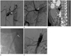
A 5-Fr RH catheter (Cook, Bloomington, IN, USA) was inserted in the right common femoral artery. Angiography confirmed CMT with significant stenosis of the trunk and complete occlusion of the proximal portion of the SMA (Fig. 1A). The distal portion of the SMA showed faint retrograde filling via the pancreaticoduodenal arcade. We first attempted to cross the obstructed SMA through the stenotic trunk, using a 5-Fr Cobra catheter (Cook, Bloomington, IN, USA) and 0.035-inch guide wire (Radifocus; Terumo, Tokyo, Japan). However, the catheter persistently slid into the connected celiac artery because the completely occluded SMA had a downward course and the ostium of the CMT had severely stenosed and could not negotiate the SMA. After several unsuccessful attempts to negotiate the SMA, we decided to first treat the CMT ostium stenosis. A 6-Fr renal guiding catheter (Cordis, Miami, FL, USA) was advanced over the wire so that the tip was placed proximal to the ostium of 460the CMT. Next, the 0.035-inch guide wire was changed to a 0.014-inch guide wire (Transend; Boston Scientific, Natick, MA, USA). A 6 × 15-mm balloon expandable stent (Palmaz; Cordis, Miami, FL, USA) was placed over the guide wire and positioned across the stenotic portion of the trunk, and the balloon was inflated. Selective angiography of the CMT revealed a patent stent and retrograde filling of the distal SMA via the pancreaticoduodenal arcade (Fig. 1B). The patient's symptoms were immediately resolved after the procedure, and she was discharged 2 days later.
One month later, she complained of recurrent abdominal pain and weight loss. Abdominal CT revealed a patent CA stent and complete occlusion of a 3.0 cm length of the proximal SMA. Retrograde flow into the distal SMA occurred via the pancreaticoduodenal arcade. No signs of ischemia, such as bowel wall thickening or abnormal contrast enhancement, were observed (Fig. 1C). Endovascular revascularization for SMA recanalization via the left brachial artery route was decided because of the downward course of the SMA and because of the difficulty in wire selection and stent placement due to prior insertion of the CA stent making the femoral approach difficult. After inserting a 6-Fr sheath, a 5-Fr Cobra catheter was inserted along the 0.035-inch guide wire. The wire was successfully negotiated through the previously inserted stent mesh into the SMA occlusion site. A 6 × 60-mm stent (S.M.A.R.T.; Cordis, Miami, FL, USA) was inserted along the wire and was carefully positioned across the obstructed SMA site. The proximal portion of the stent was overlapped in the trunk with the CA stent, and the 6-mm balloon (Synergy; Boston Scientific, Natick, MA, USA) was inflated (Fig. 1D). Final angiogram showed normal position of the overlapped CA and SMA stents and antegrade flow into the SMA and CA without residual stenosis (Fig. 1E). There were no complications, and the patient's symptoms were immediately resolved. Six months later, the patient had no clinical symptoms, and she regained her weight.
DISCUSSION
The cause of CMI is progressive atherosclerotic stenosis or occlusion of one or more mesenteric arteries (1). However, clinical manifestations of CMI are rare because atheromatous disease usually involves the proximal portion of the mesenteric arteries, allowing collateral blood flow to the intestine (2). Treatment of stenosis or occlusion of mesenteric vessels is indicated for intestinal ischemia-related symptoms, such as postprandial pain, weight loss, nausea, and diarrhea (3). The aims of treatment are to improve symptoms and prevent ischemic injury to the bowel.
Historically, CMI has been treated by surgical revascularization. However, surgery has a 15-47% morbidity rate and a 0-17% mortality rate, higher than that of endovascular treatment (1). The overall outcome of the endovascular treatment of CMI compares favorably with surgery (7). Although endovascular treatment has a higher incidence of recurrent symptoms compared with open surgery, repeat therapy is possible. Therefore, the endovascular treatment of CMI has become the first-line therapy (2, 4).
CMT, an anatomic variant where the CA and SMA have a common origin from the aorta, accounts for only 1.5% of all splanchnic artery anomalies (6). When more than one mesenteric artery is occluded or stenotic, endovascular treatment should begin with the vessel that is technically easier to access (1). Although ischemic symptoms improve when only one of the two affected arteries is successfully revascularized, treatment of the other increases bowel perfusion and prevents recurrent symptoms in the case of restenosis (3). In our case, the ostium of the CMT showed severe stenosis, and there was complete occlusion of the proximal SMA. The wire required catheter or sheath support to pass through the SMA occlusion site, but because the origin of the CMT was too stenotic, our initial attempt to access the SMA was unsuccessful. Therefore, we decided to first stent the significant stenosis of the CMT ostium, and treat the SMA occlusion later.
Another treatment option for this patient was percutaneous transluminal angioplasty (PTA) of ostial stenosis of the trunk, followed by stent placement through the occluded SMA. However, we did not choose this method, because of the risk of distal embolization of the hepatic artery and significant recoil after PTA (5). Retrograde recanalization of SMA via collaterals from the celiac artery was another treatment option. In our patient, however, SMA recanalization in retrograde fashion via the pancreaticoduodenal arcade was technically impossible because of tight stenosis at the CMT origin (8).
To our knowledge, this is the first report of endovascular treatment of CMT occlusion by crossing the two stents. Ailawadi et al. (9) reported a case of CMT occlusion treated surgically using a bypass graft. Ayers et al. (10) treated a patient with CMT occlusion using blunt microdissection catheter (Frontrunner X39 CTO Catheter; LuMend, Redwood City, CA, USA) and stent placement. However, in this case, unlike ours, the occluded CMT showed a residual normal stump that could support a catheter or sheath and enable SMA stenting.
In conclusion, in cases where SMA negotiation is technically difficult or impossible, endovascular recanalization of a stenotic CMT can be achieved by crossing the two stents. This treatment option may be used to treat CMI associated with other anatomic variations.
 XML Download