

 PDF
PDF ePub
ePub Citation
 Print
Print


INTRODUCTION
The extraocular muscle (EOM) is the most frequent site for the involvement of thyroid orbitopathy, followed in order by inferior, medial, superior, lateral rectus, and oblique muscles. The EOM is the rarest site for the involvement of extranodal marginal zone B-cell lymphoma of mucosa-associated lymphoid tissue (MALT lymphoma) in the orbit. Several cases of MALT lymphoma involving the EOM have appeared in the English literature, and most of these are in the form of case reports (1-5). The imaging findings of MALT lymphoma involving the EOM are limited to those of a single imaging modality only (1-3). As such, imaging features of this rare entity at CT, MRI, and 18F-fluorodeoxyglucose positron emission tomography/CT (18F-FDG PET/CT) altogether in a single case have not appeared in the literature.
We report here a case of MALT lymphoma involving the medial rectus muscle in a 47-year-old man along with imaging features at CT, MRI, and 18F-FDG PET/CT.
CASE REPORT
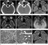
A 47-year-old man presented with a chief complaint of right ocular discomfort lasting for 1 year. He had not experienced ocular pain or diplopia. His past history was unremarkable. The ophthalmologic examination disclosed proptosis of the right eye, but the motion of the EOM was preserved. The laboratory tests, including blood cell count, biochemistry, and thyroid hormones, were normal. CT, performed by using a SOMATOM Definition Flash scanner (Siemens Healthcare, Forchheim, Germany) with a slice thickness of 3.0 mm revealed a fusiform enlargement isolated to the right medial rectus muscle, appearing as isoattenuating compared with the brain on non-enhanced CT images, and showed homogeneous, moderate enhancement on contrast-enhanced CT images, with involvement of the tendinous insertion (Fig. 1A, B). Orbital MRI was performed for better characterization of the lesion and for better assessment of the extent by using a 3.0-T unit (Signa Excite; GE Medical System, Milwaukee, WI, USA). The enlarged medial rectus muscle was isointense to the gray matter on fast spin echo (FSE) T1-weighted images (T1WI) [repetition time (TR)/echo time (TE) = 650.0/13.6; echo train length (ETL) = 3; field of view (FOV) = 160 × 160; matrix number/number of excitation = 320 × 192/2; slice thickness = 3.0 mm] with fat saturation (Fig. 1C) and FSE T2-weighted images (T2WI) (TR/TE = 5000.0/111.9; ETL = 20; FOV = 160 × 160; matrix number/number of excitation = 384 × 224/2; slice thickness = 3.0 mm) with fat saturation (Fig. 1D), and showed homogenous intense enhancement on gadolinium (Gd)-enhanced FSE T1WI with fat saturation (Fig. 1E). 18F-FDG PET/CT, performed by using Discovery STE (GE Healthcare, Milwaukee, WI, USA), demonstrated increased FDG uptake [maximum standardized uptake value (SUVmax) = 4.9 g/mL] in the right medial rectus muscle with fusiform enlargement (Fig. 1F). No abnormally increased FDG uptake was noted in other parts of the body. In view of clinical and imaging findings, our tentative diagnosis was lymphoma and euthyroid Graves' ophthalmopathy. The patient underwent biopsy of the right medial rectus muscle. The histological and immunohistochemical findings of the biopsy specimen were interpreted as MALT lymphoma (Fig. 1G, H). No findings of bone marrow involvement were documented in the bone marrow biopsy. Fractionated external beam irradiation of a total dose of 3600 cGy was applied to the right orbit. Near complete resolution of the MALT lymphoma of the right medial rectus muscle was achieved at follow-up MRI 230 days after the initial study (Fig. 1I). The patient underwent endoscopic biopsy for a raised, erosive mucosal lesion of the stomach 250 days after initial diagnosis of orbital MALT lymphoma, which revealed gastric MALT lymphoma. He underwent additional curative fractionated external beam radiation therapy at a dose of 3060 cGy targeted to the stomach. Follow-up endoscopic examination with biopsy, performed 133 days after initial diagnosis of gastric MALT lymphoma, demonstrated complete remission. No evidence of recurrent disease was observed in the orbit, stomach, or elsewhere until 13 months after complete remission.
DISCUSSION
Thyroid orbitopathy is the most common cause of abnormal enlargement of the EOM. Enlargement of the EOM associated with thyroid orbitopathy is bilateral and symmetric in 70% of cases. The inferior rectus muscle is most frequently affected, and the muscle enlargement is characteristically fusiform with sparing of tendinous insertion. The most common nonthyroidal causes of EOM enlargement are inflammatory and vascular disorders, and neoplastic diseases (6). MALT lymphoma, an extranodal, monoclonal, small B-cell proliferation, is an indolent disease that usually presents as a localized extranodal tumor. MALT lymphoma arises from various mucosal and nonmucosal tissues. It most commonly occurs in the stomach, but can involve the orbital/ocular adnexa, salivary glands, and lungs (7). Orbital MALT lymphoma most frequently involves the conjunctiva (51%), while the EOM is the rarest site of involvement (5%) (8). Several cases of MALT lymphoma involving the EOM have been presented in the English literature in the form of case reports or clinical studies (1-5). Of them, a few cases have presented imaging features (1-3). However, none have described imaging findings in detail. Rossman et al. (1) reported a case of an enlarged medial rectus muscle caused by MALT lymphoma that was misdiagnosed as thyroid orbitopathy for over 3 years, even though the patient was euthyroid. CT of the orbits simply showed enlargement of the medial rectus muscle with lateral displacement of the globe. Byard et al. (2) reported a case of MALT lymphoma isolated to a single extraocular muscle, presenting as an approximately 30 × 15-mm-sized mass arising from the region of the left superior rectus muscle with no signs of bone erosion. Benetatos et al. (3) reported a case of MALT lymphoma in which Gd-enhanced MR imaging revealed tumefaction of the inferior rectus muscle with subsequent decrease of tumor extent on follow-up study 6 months after rituximab treatment.
Our case represents the first case in which imaging findings at CT, MRI, and 18F-FDG PET/CT were presented together in a single case of this rare entity. In our case, the medial rectus muscle which involved the MALT lymphoma was manifested as a fusiform enlargement of the muscle on CT and MRI, which resembled findings of thyroid orbitopathy. However, the differentiation from thyroid orbitopathy was established by involvement of the tendinous insertion in MALT lymphoma of our case. 18F-FDG PET/CT findings of MALT lymphoma involving the EOM have not appeared in the English literature. The SUVmax of MALT lymphoma involving the medial rectus in our case was 4.9 g/mL, which was similar to that of MALT lymphoma elsewhere (9). We hypothesized that the low FDG uptake (SUVmax = 4.9 g/mL) of MALT lymphoma involving the medial rectus muscle in our case might be related to the indolent course of this entity.
Although MALT lymphomas respond well to surgical removal, radiotherapy is the treatment of choice for localized disease in the orbit/ocular adnexa. A dose of 30-35 Gy has been reported to be sufficient to provide local control and cure of the disease localized to the orbit/ocular adnexa and is associated with excellent survival (10).
In summary, it should be noted that the EOM is a rare location for the involvement of MALT lymphoma, and fusiform enlargement of EOM in MALT lymphoma may mimic that of thyroid orbitopathy on CT or MRI. The differentiation can be established by the fact that the tendinous insertion of EOM is involved in MALT lymphoma, which is not the case in the thyroid orbitopathy. In cases with solitary muscle enlargement without features of thyroid orbitopathy, muscle biopsy should be considered to exclude MALT lymphoma.
 XML Download