

 PDF
PDF ePub
ePub Citation
 Print
Print


INTRODUCTION
Ossifying fibroma is a rare, well-demarcated, fibro-osseous tumor that is composed of bone, fibrous tissue and cementum. It usually occurs in the craniofacial bones, with the mandible being the most common site. Ossifying fibroma originating from the middle turbinate is extremely rare (1, 2). Psammomatoid juvenile ossifying fibroma (PsJOF) represents a unique subtype of fibro-osseous lesion. It usually occurs in the sinonasal area of children and adolescents, and has a tendency toward locally aggressive behavior.
We report here a case of PsJOF of the middle turbinate with aggressive imaging features in an 18-year-old adolescent female along with CT, MRI and pathologic features.
CASE REPORT
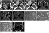
An 18-year-old adolescent female was presented with proptosis of her right eye. She had experienced proptosis and right nasal obstruction for 3 years. On physical examination, a large mucosa-lined mass was identified in the right nasal cavity. No palpable mass elsewhere or cervical lymphadenopathy was identified. Multidetector CT showed that an approximately 35 × 42 × 41 mm-sized, well-demarcated, expansile, heterogeneous mass originated from the right middle turbinate and occupied the right nasal cavity and ethmoid sinus (Fig. 1A-C). The tumor matrix consisted of areas of ground-glass attenuation with moderate enhancement, non-enhancing cystic spaces and irregular ossifications (Fig. 1B, C). It was surrounded by a thick peripheral rim ossification (Fig. 1B, C). Focal bony destruction was noted at the right lamina papyracea and cribriform plate (Fig. 1A). MRI was performed for better characterization of the mass. T1-weighted images (T1WI) portrayed that the mass was heterogeneous and hypointense compared with the brain (Fig. 1D). T2-weighted images (T2WI) with fat saturation revealed heterogeneous mass containing multiple hyperintense cystic spaces (Fig. 1E), which corresponded to the non-enhancing, hypointense areas within the moderately enhancing tumor matrix on gadolinium (Gd)-enhanced images (Fig. 1F). In view of CT and MR imaging features, our tentative diagnosis was ossifying fibroma, and alternative diagnoses included fibrous dysplasia, osteosarcoma and metastatic diseases. The histological examination of the specimen obtained by endoscopic sinus surgery (ESS) confirmed the diagnosis of PsJOF. Histologically, the lesion consisted of highly cellular and loose or myxoid areas (Fig. 1G). It was characterized by the presence of small mineralized (psammomatoid) bodies admixed with intervening fibroblastic stroma (Fig. 1H). The trabeculae were characteristically lined by osteoblastic rimming (Fig. 1I).
The postoperative course was uneventful, and there has been no evidence of tumor recurrence until 13 months after surgery. A follow-up bone algorithm non-enhanced CT of the sinonasal area 13 months after ESS revealed some residual disease of ground-glass attenuation at the right medial orbital wall and cribriform plate (Fig. 1J), the areas that could not have been easily approached during ESS. The patient is scheduled to undergo a second ESS for residual disease under the guidance of a CT-based navigation system.
DISCUSSION
Fibro-osseous lesions are rare in the sinonasal area (1-3). Fibrous dysplasia and ossifying fibroma are included in this category. Histologically, the lesion of ossifying fibroma reveals trabeculae composed of lamellar and woven bones, variable amounts of vascularized fibrous stroma, and osteoblastic rimming of the trabeculae. In addition, there may be areas of cementum that appear as psammoma bodies embedded in fibrous stroma (4-6), while the lesion of fibrous dysplasia shows irregularly shaped trabeculae composed of woven bone only; moreover, there is no osteoblastic rimming. The fibrous stroma is often less vascular and cellular than that in an ossifying fibroma.
Ossifying fibroma is typically a benign, slowly growing neoplasm; however, cases showing aggressive behavior and rapid growth have been reported. Juvenile ossifying fibroma, which can be classified into two distinct histopathologic variants (psammomatoid and trabecular), is benign (however, aggressive neoplasm is commonly seen in children below 15 years of age) and has a high tendency for recurrence. PsJOF, characterized histologically by the presence of small mineralized (psammomatoid) bodies admixed with a cellular stroma, varying amounts of myxomatous material, and scattered giant cells, occurs predominantly in the sinonasal and orbital bones, while trabecular juvenile ossifying fibroma, characterized by trabeculae of fibrillary osteoid and woven bone, predominantly affects the jaw.
Regarding the behavioral differences, ossifying fibroma is known to enlarge even after skeletal growth ceases, whereas fibrous dysplasia usually stops progression after puberty (6, 7). Accordingly, ossifying fibroma should be resected whenever possible, although "wait and see" with regular clinical and imaging follow-up is appropriate for fibrous dysplasia (8, 9). As such, a correct diagnosis is prerequisite for appropriate management. However, owing to the overlapping clinical and histomorphologic features of fibro-osseous lesions, the diagnosis of fibro-osseous lesions of the head and neck is not always easy. It is often difficult to diagnose them histologically. Thus, the correlation of imaging and pathologic findings is prerequisite for establishing a definitive diagnosis.
Radiographically, the lesion of an ossifying fibroma is well-demarcated from the surrounding bone by a radio-opaque border, whereas fibrous dysplasia is presented with a diffuse border. The CT features of a typical ossifying fibroma consist of a monostotic and well-demarcated lesion, which contains tissues of varying attenuation values, while those of fibrous dysplasia are poorly delineated osseous expansion covered by a thin cortex. Han et al. (10) reported CT and MR imaging findings of five case of sinonasal psammomatoid ossifying fibroma, four in the sphenoethmoidal area with extension into the nasal cavity or orbit and one in the perpendicular plate of the ethmoid bone. They noticed that the lesion was well-circumscribed, multiloculated, and expansile with a thick wall of bone density on CT, and showed enhancement on postcontrast MR images. Ossifying fibroma of the nasal turbinates is extremely rare. Only four cases have appeared in the English literature (1-4). Caylakli et al. (1) reported a case of ossifying fibroma that showed a monostotic, hyperdense (ground-glass attenuation) lesion confined to the middle turbinate. Galvan et al. (2) reported a case of ossifying fibroma that showed a large, expansile mass arising from the left middle turbinate and extending to the ethmoid sinus with an extensive ground-glass attenuation surrounded by hypoattenuating fibrous tissue and/or cystic areas on CT. The ground-glass attenuation area on CT corresponded to a well-defined, intermediate to slightly low signal intensity area with homogeneous enhancement on T1WI and Gd-enhanced T1WI, and a low signal intensity area on T2WI. CT and MR imaging findings of the cases of Caylakli et al. (1) and Galvan et al. (2) are substantially different from those of the present case in that the tumor matrix of our case consisted of areas of ground-glass attenuation, non-enhancing cystic spaces and irregular ossifications. Furthermore, the lesion of our case demonstrated moderate solid enhancement and aggressive imaging features, including bony erosion or destruction of the lamina papyracea and cribriform plate. As such, our case presents a diagnostic dilemma because these imaging findings may be seen in various benign and malignant sinonasal diseases, such as fibrous dysplasia, osteosarcoma and metastatic diseases. However, the long clinical history of disease and the expansile growth of the lesion in our case favor benign entities. Recurrence or residual disease may be attributed to incomplete resection, due to the location of the lesion as in our case, and the infiltrative nature of the tumor borders.
In summary, our case represents an extremely rare PsJOF originating from the middle turbinate, demonstrating various tissue components by CT and MRI. PsJOF of the middle turbinate may be presented with a well-demarcated, expansile, solidly enhancing mass with focal bony destruction, which may mimic various benign and malignant neoplasms of the sinonasal area. A combination of clinical, imaging and histological features enables establishing an accurate diagnosis of PsJOF.
 XML Download