

 PDF
PDF ePub
ePub Citation
 Print
Print


INTRODUCTION
The greater omentum is a double-layered peritoneal structure composed mainly of fatty tissue and serpentine gastroepiploic vessels. It attaches to the greater curvature of the stomach and the proximal part of the duodenum, covering the small bowel and reflecting at the level of the pelvic inlet toward the transverse colon like an apron (1). Omental infarction is an uncommon cause of right lower quadrant (RLQ) pain in children who visit the emergency room. Although relatively common in adults, it is rare in children, who account for only 15% of cases (2). Helmrath et al. (3) reported just 18 cases of omental infarction in 15 years of experience.
The etiology of omental infarction is unknown. Embryonic vascular variants of the omentum with vascular kinking or torsion of the right epiploic vein may cause omental infarction with RLQ pain (4-6). Thrombosis of an omental vessel due to a heavy meal or obesity can also induce omental infarction (7, 8). Rarely, omental infarction occurs after blunt abdominal trauma such as a bicycle handlebar injury (9).
It is important to differentiate medically treatable omental infarction from other diseases that cause RLQ pain. Omental infarction is often confused with surgical conditions such as acute appendicitis or intussusception, as well as with medical conditions such as mesenteric lymphadenitis or acute appendagitis (7). The signs and symptoms of these diseases are similar and include RLQ pain, abdominal tenderness, and vomiting. Laboratory parameters, such as white blood cell (WBC) counts, erythrocyte sedimentation rate (ESR), C-reactive protein (CRP), or other inflammatory findings, are also not diagnostic (7). Therefore, it is important to differentiate it from other conditions to avoid unnecessary surgery by using imaging modalities (10).
The purpose of this study was to evaluate the CT scan and ultrasonography (US) findings of omental infarction in children with RLQ pain and to compare them with those of other diseases that may cause similar RLQ pain and require surgical rather than medical treatment.
MATERIALS AND METHODS
All CT and US image reports from patients under 17 years old between January 2005 and March 2012 were reviewed by use of the Picture Archiving and Communication System and nine archived children with confirmed omental infarction clinically or surgically were found. Three patients who presented with severe abdominal pain and elevated WBC counts underwent appendectomy and omentectomy; the other six were treated conservatively. US and Doppler examinations were performed using a IU21, IU22 US and HDI 5000 with a 5- to 12 MHz linear array and a 4- to 9 MHz convex transducer (Philips Medical Systems, Bothell, WA, USA) and a LOGIQ E9 with a 6- to 15 MHz linear array and a 2.8- to 5 MHz convex transducer (GE Healthcare, Milwaukee, WI, USA). CT studies were carried out using three MDCT (SOMATOM Sensation 16, Siemens AG, Forchheim, Germany; Lightspeed VCT, GE, Milwaukee, WI, USA; iCT, Philips, Bothell, WA, USA) with IV contrast injection. Based on previous studies (4-6), omental infarction was defined with findings as follows: on CT, heterogenous enhancing mass-like or fatty lesion between the anterior abdominal wall and ascending colon; on US, heterogeneously aggregated fatty mass at similar position as CT; no other pathologic findings such as appendicitis or diverticulitis. If other pathologic conditions could not be ruled out, explorative laparotomy was done for further evaluation. All images were interpreted by two pediatric radiologists who had 10 and 23 years of experience. We also reviewed clinical symptoms, such as abdominal pain, presence of fever, and nausea and/or vomiting. Laboratory findings such as WBC counts, ESR, and CRP were also assessed.
RESULTS
Clinical Presentation
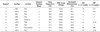
Clinical and laboratory data of the nine patients are summarized in Table 1. There were eight boys and one girl. The mean age was 8.4 years (range, 4-11 years). Presenting symptoms included RLQ pain (n = 6), epigastric pain (n = 1), right upper quadrant (RUQ) pain (n = 1), and periumbilical pain (n = 1). The mean duration of symptoms was 2 days (range, 1-4 days). No patient presented with fever (mean body temperature 36.9℃, range, 36.7-37.4℃). Six of nine patients showed increased WBC counts (> 10000 × 103/µL). Five patients tested ESR count and CRP, and five patients had an elevated ESR count and three had an elevated CRP.
Imaging Interpretation
CT Findings
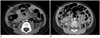
The CT findings of the nine patients are summarized in Table 2. Seven patients had heterogeneously enhanced mass-like lesion between the ascending colon and the abdominal wall, located in the RLQ (Fig. 1A) in six patients and in the RUQ in one. The lesions were triangular fatty mass in six patients, an oval fatty mass in one. Among these patients, two had rim enhancement around fatty mass. The remaining two patients had only ill-defined diffuse fat infiltration.
US Findings
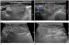
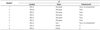
Three patients underwent abdominal US with both gray scale and color Doppler imaging, the findings of which are summarized in Table 3. All three cases had an ill-defined heterogeneously aggregated fatty mass in the RLQ. Two of these had decreased vascularity of the heterogeneously echoic RLQ mass with color Doppler imaging. The vascularity of the fatty mass was increased in one case, but it had decreased by 1 day later (Fig. 2).
Treatment and Follow-Up
Three patients underwent surgery, at which point omental infarction was confirmed. One patient was diagnosed with omental infarction by ultrasonography and was treated conservatively. One day later, the patient complained of aggravating abdominal pain. His WBC counts were elevated and neutrophil differentiation was noted. On follow-up ultrasonography, the appendix could not be delineated, so laparoscopic appendectomy and omentectomy was performed to differentiate between perforated appendicitis and omental infarction. Two other patients presented with persistent pain, tenderness and rebound tenderness in the RLQ. With these two patients, appendix was not clearly delineated, so acute appendicitis could not be completely ruled out. Laparoscopic appendectomy and omentectomy were performed and omental infarction was pathologically confirmed. Their appendices were normal.
Two patients had follow-up CT or US. In one patient, the size and enhancement of omental fat infiltration in the RLQ on CT was decreased after 1 month (Fig. 1B). Another patient underwent follow-up US after 1 month, and the fat infiltrations in the RLQ had disappeared. The remaining four patients were completely relieved from abdominal pain after conservative treatment; they had no surgical or imaging follow-up.
DISCUSSION
Omental infarction is uncommon in children with RLQ pain, with more than 85% of reported cases occurring in adults (4). Its etiology is unclear, but omental infarction can occur with or without omental torsion. It is associated with several predisposing factors, such as obesity, venous congestion after a large meal, vascular kinking, and rarely, blunt trauma (4-9). Left-sided acute omental infarction is far less common than right-sided (8, 11).
The typical CT appearance is a solitary, well-defined, triangular or ovoid mass between the abdominal wall and transverse or ascending colon that has a heterogeneous, sometimes whorled pattern of linear fat strands, and surrounding fat infiltration may be present (4). In this study, the CT findings were typical, a mass between the abdominal wall and ascending colon of the RLQ in six patients and a similar lesion of the RUQ in one.
On US, omental infarction appears as a solid, non-compressible, painful and moderately hyperechoic mass near other normal abdominal structures. The lesion is typically found in the RLQ, just under the abdominal wall overlapping the right colon (4, 5). In our study, two patients had a heterogeneous ill-defined fatty mass in the RLQ and one in the RUQ, located between the abdominal wall and the right colon.
On Doppler ultrasonography, two findings indicative of omental infarction are reported. One is a hyperechoic mass containing poorly defined nodular or linear hypoechoic areas with few vessels within the mass and hyperemia in the peripheral area. On pathology, the hypoechoic area is suggestive of hemorrhagic infarction (6). The other finding is a hyperemic hyperechoic mass that contains an avascular hypoechoic tubular structure (8). In this study, three patients underwent color Doppler ultrasonography and two had decreased vascularity, suggesting infarction. The other patient initially presented with a hyperemic hyperechoic mass, but the vascularity was decreased in the follow-up study, suggesting progression of the infarction.
Three patients underwent surgery due to misdiagnosis of acute appendicitis, and all three underwent appendectomy and partial omentectomy. The omentum was an unusual color grossly, and exhibited infiltration of inflammatory cells microscopically. Hemorrhagic congestion and inflammatory cell infiltration are known pathologic findings of omental infarction (4).
Symptoms of omental infarction are similar to other diseases such as acute appendicitis, acute appendagitis, mesenteric lymphadenitis, diverticulitis, and infective enterocolitis. Acute appendicitis is one of the most common emergency causes of RLQ pain. Differentiation of these conditions by clinical and laboratory findings is difficult (9, 12). Yang et al. (13) reported that patients with omental infarction show less fever, nausea, vomiting, lower WBC and neutrophil counts, and lower CRP values, than those with acute appendicitis. CT criteria for acute appendicitis are an enlarged appendix with a diameter of more than 6 mm, or a wall thickness of more than 3 mm, in conjunction with periappendical inflammatory changes. The sensitivity and specificity of CT for acute appendicitis have been reported to be 94-98% (14).
Epiploic appendagitis is defined as inflammation of the epiploic appendage, which is a round, fat-containing peritoneal pouch, prominent around the sigmoid and descending colons. Therefore, appendagitis typically manifests as LLQ pain and can mimic acute diverticulitis. Typical CT imaging shows a pericolic oval fatty attenuated lesion with an enhanced internal dot and peripheral rim, and surrounding fat infiltration. Adjacent colonic wall thickening is sometimes noted. As mentioned previously, omental infarctions are usually localized to the right lower quadrant and lack the hyperattenuated rim, features which aid diagnosis (15-17). If acute appendagitis occurs at the right side of the abdomen, it usually located at pericolic region, oval-shaped, and smaller than an infracted greater omentum. In addition, omental infarction does not show central dot which can be found on epiploic appendagitis. Clinically these two diseases are self-limited condition and tend to resolve spontaneously, so it is less important to differential diagnose these two diseases (17).
Mesenteric lymphadenitis is relatively frequent in children and is a diagnosis of exclusion (18). There are three or more clustered lymph nodes around the small bowel mesentery and anterior to the psoas muscle. On CT, right-sided mesenteric lymphadenopathy in the absence of other inflammatory conditions is a characteristic finding (19). On US, features indicative of mesenteric lymphadenitis are enlarged mesenteric lymph nodes (over 4 mm in diameter), associated mucosal thickening of the terminal ileum, and lack of other diseases such as appendicitis (20).
Diverticuli typically affect the left lower abdomen, due to the sigmoid colon being most commonly affected. The right colon and rectum are less often affected, and both usually cause RLQ pain. CT findings of acute diverticulitis include asymmetric or circumferential colonic wall thickening with focal pericolic fat infiltration. Colonic diverticuli with inflammation and abscess formation are also noted. Typically, acute diverticulitis is seen in older patients more commonly than is omental infarction, and presents with nausea, vomiting, elevated leukocyte counts, diffuse abdominal pain and/or rebound tenderness (19). The differentiation of acute diverticulitis from omental infarction by identifying inflamed diverticuli under CT or US scan. The mesenteric fat infiltration induced by acute diverticulitis is commonly much extensive than that induced by epiploic appendagitis or omental infarction (17).
Infectious colitis is relatively common, and manifests as acute abdominal pain. It can be indistinguishable from omental infarction or acute appendicitis, particularly when the cause is infection of the ileocecal region by Yersinia enterocolitica, Campylobacter jejuni, or Salmonella enteritidis (19).
This study had several limitations. First, the study population was relatively small. However, omental infarction is uncommon in children and for this reason, most previous studies also involved small groups of patients. Second, although, omental infarction was treated conservatively, only three cases were pathologically confirmed.
In summary, patients with omental infarction presented with RLQ pain and tenderness. The lesion was most commonly located between the abdominal wall and right colon. Typical CT findings were a well-defined, heterogeneously enhanced triangular or ovoid fatty mass. A solid, non-compressible, hyperechoic mass was the typical US finding. Vascularity is decreased as the disease progresses on color Doppler, which is compatible with the general pathology of infarction.
In conclusion, it is important to be familiar with the typical imaging features of omental infarction to facilitate its differentiation from other conditions that present with RLQ pain.

 XML Download