

 PDF
PDF ePub
ePub Citation
 Print
Print


INTRODUCTION
Anterior chest wall pain and swelling associated with inflammatory symptoms may be related to the inflammatory process such as infections and arthritis (1). Occasionally, an underlying pathologic process, such as malignancy, can be missed if the symptoms clearly implicate an infection. It can lead to catastrophic results for both the patient and the clinician. Here, we report an elderly male patient initially presenting as gas gangrene in the pectoralis major muscles and osteomyelitis of the sternum and finally diagnosed as metastatic carcinoma of unknown origin of the sternum.
CASE REPORT
A 69-year-old man was admitted for evaluation of fever and pain in his anterior chest wall. He had visited several orthopedic clinics for the anterior chest wall pain for 3 months, but the symptoms were not improved and progressing. On admission, his body temperature was 39.8℃. The pain was centered on the anterior chest wall, especially around the sternum. The soft tissue of the anterior chest wall was swollen, and the overlying skin was mildly erythematous with local heating sensation. On palpation, severe pain with crepitus was present. The remainder of the physical examination revealed only the left-sided hemiplegia due to a previous cerebral infarction occurred 10 years ago, and he had no history of diabetes.
A complete blood cell count showed a hemoglobin level of 13.0 g/dL, 21900/mm3 leukocytes with 88% neutrophils and 4.4% lymphocytes, and a platelet count of 240000/mm3. The erythrocyte sedimentation rate (ESR) was 121 mm at the first hour, and the C-reactive protein (CRP) was 30 mg/L (normal < 0.7). The aspartate transaminase was elevated at 98 IU/L (normal < 35 IU/L), but the alanine transaminase level was 39 IU/L (normal < 35 IU/L). The gamma glutamyl transpeptidase was 75 IU/L, within the normal range. The blood urea nitrogen and creatinine levels were within the normal range. The serum lactate dehydrogenase was elevated at 643 IU/L (normal < 450 IU/L). The serum total protein was 5.6 g/dL with a serum albumin of 2.2 g/dL. The serum alkaline phosphatase was 252 IU/L (normal < 126 IU/L). The serum creatine phosphokinase and myoglobin were both elevated at 309 (normal < 100 IU/dL) and 149.0 ng/mL (normal < 70 ng/mL), respectively. The blood culture was negative.
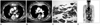
On the initial contrast-enhanced chest CT scan (Brilliance 64; Philips Medical Systems, Cleveland, OH, USA), there was a cortical disruption in the sternum, and air bubbles were also seen in the sternal marrow and retrosternal space. Gas was bilaterally spread throughout the pectoralis muscles to the axilla (Fig. 1A). The soft tissue surrounding the sternum was diffusely enhanced from the level of the 2nd costosternal junction to the 5th. Especially, at the level of the 3rd costosternal junction, lobular enhancing soft tissue masses were suspected anterior to the sternum (Fig. 1B). We suggested sternal osteomyelitis with gas gangrene. Additionally, the CT images demonstrated peripheral consolidation in both lower lobes and a minimal amount of bilateral pleural effusion, thought to be due to pneumonia. However, the whole lung evaluation using the lung window settings revealed no definable lung masses or nodules.
Fine needle aspirations for the pectoralis muscle and sternal marrow were performed with Gram staining and both the aerobic and anaerobic culture. Empirical intravenous antibiotics were then initiated (ceftriaxone, clindamycin, and gentamicin). All Gram stains and cultures from the aspirates were negative. The polymerase chain reaction was negative for both tuberculosis and non-tuberculous mycobacteria. Fungal cultures were negative. Even after a 2 week-course of empirical antibiotics and analgesics, the soft tissue swelling on the anterior chest wall, especially around the sternum, did not regress. A follow-up CT scan was then obtained.
On the second contrast-enhanced CT scan, the previously observed extensive gas through the pectoralis muscles had resolved. However, the enhancement of soft tissue surrounding the sternum remained without change. An incisional biopsy for the patient's sternum and the suspicious soft tissue masses was performed. The histopathologic results were consistent with metastatic, poorly-differentiated carcinoma, most likely squamous cell carcinoma (Fig. 1C).
Then, the patient underwent a work-up looking for the primary malignancy. A whole body positron emission tomography (PET)-CT scan was performed and demonstrated multiple increased uptakes suggesting bone metastases to the axial skeleton, including the spine, rib, pelvis, and skull base, especially the clivus (Fig. 1D). Pelvic nodal metastases around the prostate were also revealed. In addition, brain MRI was performed to evaluate the skull base. A metastatic mass involved the clivus, but there was no evidence of a primary tumor in the nasopharynx.
Among the serum tumor markers, serum prostate specific antigen (PSA) was 17.33 ng/mL (normal < 4 ng/mL), but special staining of the specimens from the sternum and the surrounding soft tissue were all negative for PSA. Other tumor markers, including chorioembrionic antigen 19-1, were all within the normal range.
The patient refused prostate biopsy and any further evaluation. He also refused treatment except for pain control. The chest wall masses gradually enlarged, and his pain became multifocal. His condition declined, and he expired 2 months after the diagnosis due to multiple organ failure.
DISCUSSION
When inflammatory skin changes are associated with malignancy, these conditions are named according to the malignancy, such as inflammatory cancer, inflammatory carcinoma, and inflammatory metastasis. In our patient, the soft tissue swelling and pain of the anterior chest wall and erythematous skin with local heating sensation were due to the sternal metastasis. Moreover, cortical disruptions of sternum resulted in gas spreading through the pectoralis muscles. We assumed this condition as inflammatory metastatic carcinoma of sternum. Bachmeyer et al. (1) also described this condition as 'inflammatory sternal metastasis' in their report of two patients with sternal metastasis from lung adenocarcinoma. The patients had fever above 38℃ and elevated ESR and CRP level, similar to our case. There was another case of inflammatory sternal metastasis from a squamous cell carcinoma reported by Thongngarm et al. (2). They reported a 44-year-old woman who presented with pain in the upper sternum and costochondral junction. At first, the patient was diagnosed with Tietze's syndrome and was treated with the non-steroidal anti-inflammatory drugs. As palpable tenderness and mild swelling of the anterior chest wall developed, fine needle aspiration was performed. The pathology revealed squamous cell carcinoma of unknown origin. The different findings between the three previous cases (1, 2) and our case were the CT findings of air spreading through the pectoralis muscles. We first considered these lesions as gas gangrene associated with osteitis and osteomyelitis of sternum and costochondral junction. The possibility of a malignancy arose after the follow-up CT demonstrated no regression of the soft tissue enhan-cement despite the resolution of the intramuscular gas.
An interesting finding in the current case is that the spontaneous gas gangrene was associated with a sternal metastasis, and this is the first such association to be reported to our knowledge. Usually, gas gangrene is caused by the clostridium species such as Clostridium perfringens or a polymicrobial infection and associated with a contaminated penetrating muscle injury. In our case, no microbe was isolated on culture, and there was no obvious wound to suggest a route of entry. Moreover, gas gangrene is a rapidly progressive and very fatal condition involving all the muscles, subcutaneous, and skin layers, and requires urgent surgical debridement (3, 4). Our patient, though, did not show the usual deteriorating clinical course of gas gangrene.
We attempted to find the primary malignancy. The sternum is not a common site for the involvement with malignancy regardless of primary or metastatic lesion. The common malignancies of the sternum are metastatic adenocarcinoma from the breast, thyroid, kidney, gastrointestinal tract, liver, lung, and rarely, the prostate (2, 5, 6). In our patient, the bone metastases were already too extensive, as was apparent on the PET-CT scan. The only positive tumor marker was the serum PSA, but the histopathology suggested squamous cell carcinoma, not adenocarcinoma. Considering that both the prostate and breast cancer can undergo squamous transformation, we performed the special staining for PSA on the sternal tissue, which turned out to be negative. Ultimately, we could not identify the primary site of the metastatic squamous carcinoma of sternum during the patient's life.
It is common and reasonable for physicians to consider a painful swelling of the anterior chest wall as a benign condition including infection, inflammatory arthritis, relapsing polychondritis, seronegative spondyloarthropathies, and crystalline-induced arthritis (2, 6, 7) and to develop the treatment plans accordingly. An inflammatory sternal metastasis is a rare condition reported in few reports. Here, we emphasize that the metastasis of the sternum and adjacent costochondral junction may present with inflammation. The definite diagnosis should also depend on the histopathological examination and bacteriologic studies.
 XML Download