

 PDF
PDF ePub
ePub Citation
 Print
Print


INTRODUCTION
Phyllodes tumors are defined as a group of circumscribed biphasic tumors, basically analogous to fibroadenomas, characterized by a double-layered epithelial component arranged in clefts surrounded by an overgrowing hypercellular mesenchymal component, typically organized in leaf-like structures (1). Phyllodes tumors are uncommon in the breast; they account for 0.3-1% of all primary tumors of the breast, and 2.5% of all fibroepithelial tumors of the breast (1). Carcinoma in a phyllodes tumor has been reported to occur in 1-2% of phyllodes tumors, and ductal carcinoma in situ (DCIS) arising in a phyllodes tumor is even more unusual (2, 3). Here, we report a case of DCIS arising in a benign phyllodes tumor.
CASE REPORT
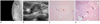
A 42-year-old woman was presented with an ovoid mass detected on a mammography (Fig. 1A). Mammography showed a 2.2 × 1.4 cm sized, ovoid, circumscribed, and iso-dense mass in the upper portion of the right breast. There was no calcification in the mass. She had no history of breast abnormalities, previous breast surgery or radiation therapy. A physical examination revealed a 2 cm ill-defined mass in the right upper outer breast. There was no overlying skin abnormality or palpable axillary or supraclavicular lymph nodes. A breast ultrasound (US) was performed to localize the lesion and for a biopsy. The breast US showed a 1.9 cm ovoid, partially microlobulated, partially circumscribed, and isoechoic mass at the 10 o'clock direction in the right breast (Fig. 1B). Doppler US showed vascularity in the mass. We classified the mass as Category 4 according to the Breast Imaging Reporting and Data System.
We then performed a US-guided core needle biopsy, and the histopathological examination of the biopsy specimen revealed several ducts with atypical ductal hyperplasia, with combined DCIS not being able to be excluded. Therefore, surgical excision was performed, and the gross specimen showed a white, firm, well-circumscribed, round mass measuring 1.8 cm in its greatest dimension. There was no intratumoral necrosis. Histologically, the well-defined tumor showed a mildly cellular stroma and an epithelial-lined cleft, and exhibited a leaf-like pattern (Fig. 1C). The stromal component revealed a mild cytologic pleomorphism, and two mitoses per 10 high power fields. These findings were consistent with a benign phyllodes tumor. Additionally, 1.2 × 1.0 × 0.9 cm sized cribriform DCIS was identified within the phyllodes tumor (Fig. 1D). Although columnar cell change with focal mild atypia appeared in the superior resection margin of the specimen, the final diagnosis was DCIS in a benign phyllodes tumor.
The patient received local radiotherapy to her right breast, and she remains well without signs of local recurrence or distant metastases on breast US and mammography 8 months following her surgery.
DISCUSSION
Phyllodes tumors are graded into benign, borderline and malignant categories, based on the histological parameters of stromal hypercellularity, cellular pleomorphism, mitotic activity, nature of the margins, and stromal pattern (1). As was seen in our patient, the features of a benign phyllodes tumor are mild or moderate stromal hypercellularity, little cellular pleomorphism, few mitoses or pushing margins, and no stromal overgrowth. Malignant transformation of phyllodes tumors usually occurs in the stromal component, and it is rare in the epithelial component. Most phyllodes tumors have a benign ductal component, but carcinoma has been reported to occur in 1-2% of phyllodes tumors, many of which are lobular carcinoma in situ (2). DCIS arising in a phyllodes tumor is very rare. We found six cases of DCIS arising in a phyllodes tumor without invasive ductal carcinoma documented in the English language medical literature (3-8). The size of the previously reported phyllodes tumors that harbor DCIS ranged from 3.5-19 cm. However, in our case, the tumor's largest dimension was 1.8 cm. This is the smallest case to have been reported. All the reported cases of DCIS within a phyllodes tumor, including the one in this case, show three benign stromal components, one borderline stromal component, and three malignant stromal components. It seems that there is no relationship between the stromal components and the occurrence of DCIS in a phyllodes tumor. It is known that the stromal components of phyllodes tumors, which harbor invasive carcinoma, are usually benign or of low grade malignancy (2).
Carcinoma arising within a phyllodes tumor is rare with less than 30 cases reported in the literature (9). The reported subtypes include in situ and invasive ductal and lobular carcinoma, tubular carcinoma and squamous cell carcinoma. The pathogenesis of the carcinoma in the phyllodes tumor is controversial. Some authors consider the epithelial component of the phyllodes tumor to have been affected by systemic growth factors and hormones, and therefore the carcinoma to have originated within the phyllodes tumor (4, 7). In such case, the carcinoma is caused by accident in the phyllodes tumor. Other researchers think that the carcinoma starts in the mammary gland near the phyllodes tumor (4). Another group proposes a third theory: that the epithelial change has been induced by the stromal component of the phyllodes tumor (4).
In most cases, it is very difficult to preoperatively detect the presence of carcinoma within a phyllodes tumor. This is due to the fact that the phyllodes tumor usually takes up a larger area than the carcinoma. In many cases, the existence of a combined carcinoma is noted for the first time, only after a postoperative histological search has been performed (7). When handling an extirpated phyllodes tumor, it is important to conduct a thorough histopathological search, always keeping in mind the possibility of a combined carcinoma. In our case, the malignant epithelial component was not detected at preoperative core needle biopsy.
We found two cases of radiologic finding of DCIS, arising in a phyllodes tumor documented in the English language literature. Nomura et al. (7) reported that mammography revealed a well-defined tumor with a diameter of 3.3 cm, and US revealed a tumor with a regular border and low echogenicity. Yamaguchi et al. (8) described the tumor as a mixed cystic and solid mass on US and magnetic resonance imaging. There are no specific clinical or radiologic clues to indicate the presence of in situ carcinoma within a phyllodes tumor (2). It is able to be detected on a mammography when the DCIS is associated with calcification. However, in our case, the tumor was detected on a mammography as a noncalcified ovoid mass.
As carcinoma within a phyllodes tumor is very rare, treatment and follow-up of these cases are not standardized. It is recommended that the treatment be customized in each case based on whether there is an invasive component, affected lymph nodes or distant metastases, and that the carcinomatous component be treated independently of the phyllodes tumor (7, 10). Axillary lymph node dissection is not part of the standard treatment for phyllodes tumors as lymph node spread is rare. Also, lymph node metastases associated with a carcinoma, within a phyllodes tumor, are extremely rare; to our knowledge, only two cases have been reported (9, 10). In our patient, no abnormal lymph node was noted in either axilla on an ultrasonography. Further, after surgical excision, postoperative radiotherapy was given only to the right breast.
The prognosis for cases of carcinoma within benign phyllodes tumors is generally favorable; however, the prognosis for a malignant phyllodes tumor may be more guarded (10). There have been no comprehensive reports concerning its prognosis up until now.
In conclusion, we report mammographic and US findings of DCIS arising within a benign phyllodes tumor. There are no mammographic or US findings suggestive of associated DCIS within a phyllodes tumor.
 XML Download