

 PDF
PDF ePub
ePub Citation
 Print
Print


INTRODUCTION
Malignant fibrous histiocytoma (MFH) is the most common soft-tissue sarcoma that occurs in adults. MFH arising from the genitourinary system is extremely rare (1), and to our knowledge, only 1 case of MFH of the ureter has been reported in Taiwan (2).
Dioctophyma renale (D. renale, the giant kidney worm) infection, an uncommon parasitic infection, has been reported to occur in various countries. This species lacks host specificity and can infect many mammalian species, including humans, although only rarely. Less than 20 cases of dioctophymatosis have been confirmed worldwide. In these cases, worms were found in various body parts such as the kidneys, peritoneal cavity, and subcutaneous layer (3, 4).
Here, we report the first case of simultaneous occurrence of primary MFH arising from the ureter and D. renale infection.
CASE REPORT
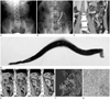
A 57-year-old man presented with pain in the left lower quadrant that persisted for 10 days. His medical history and the results of his physical examination were unremarkable. Results of laboratory tests showed microscopic hematuria (5-9 red blood cells per high-power field). Abdominal radiograph was unremarkable. Intravenous urography showed complete obstruction of the upper left ureter at the level of the upper endplate of the L3 vertebral body (Fig. 1A). Retrograde pyelography (RGP) revealed an intraluminal filling defect indicating an irregularly shaped polypoid lesion at the level of the upper endplate of the L4 vertebral body (Fig. 1B). A gap of 1 vertebral height was observed between the obstruction level identified by intravenous pyelogram and the filling defect level identified by RGP. CT scan showed a persistent, enhancing well-defined polypoid mass-like lesion of size approximately 1.0 × 1.3 × 1.7 cm, in the upper left ureter. Periureteral infiltration was not evident (Fig. 1C). The CT scan also revealed mild hydronephrosis and hydroureterosis. Diagnosis of a benign lesion, such as fibroepithelial polyp or papilloma, was suspected on the basis of this finding, which correlated with the RGP finding. However, we also suggested urothelial carcinoma as a second diagnosis. Despite our recommendation, the patient refused to undergo an operation.
Approximately 10 months after the first presentation, gross hematuria and a worm-like foreign body (approximately 12 × 0.5 cm) in the urine (Fig. 1D) were observed. A follow-up CT scan showed that the size of the lesion in the upper left ureter increased (approximately 2.3 × 2.2 × 4 cm) and that the lesion extended to the renal pelvis and to the more distal part of the ureter. The mass demonstrated a progressive enhancement pattern. Irregular wall thickening and enhancement of the left ureter were observed around the mass lesion along with periureteral and peripelvic fat infiltration. Compared to the previous CT scan, the follow-up CT scan showed a few conglomerate left paraaortic lymph nodes that had increased in size and number (Fig. 1E). In addition, a decrease in the size of the left kidney, deterioration of excretory function, and progression of hydronephrosis were observed. These findings were highly suggestive of a malignant lesion such as urothelial carcinoma.
The worm-like foreign body in the urine was confirmed to be a parasite. After 1 month, we performed a left radical nephroureterectomy. The tumor extended into the major calyx, pelvis, and proximal ureter. The kidney parenchyma was grossly unremarkable (Fig. 1F). Microscopic examination showed that the tumor was mostly composed of sheets of bizarre cells with abundant eosinophilic cytoplasm and lymphoplasma cells. Numerous epithelial-lined cysts and entrapped tubules were scattered throughout the tumor, which was lined by simple or stratified cuboidal to columnar cells with eosinophilic, granular cytoplasm. Immunohistochemical staining showed that the tumor cells were positively stained for CD68 (Fig. 1G). Ultrastructural examination showed that the tumor contained highly cellular spindle cell areas and pleomorphic cells with oval and irregular nuclei. The cytoplasm of the tumor cells consisted of rough endoplasmic reticulum, mitochondria, and some nuclei having prominent nucleoli. The three regional lymph nodes were also dissected and there was no evidence of metastasis.
The final diagnosis was that of MFH. The worm-like foreign body in the urine was confirmed to be D. renale (giant kidney worm). Distant metastasis was not evident on positron emission tomography/CT scan, and the patient was in a disease-free state in 7 months. Moreover, the paraaortic lymph nodes were decreased in size on the follow-up CT.
DISCUSSION
In 1963, MFH was first described as malignant histiocytoma and fibrous xanthoma by Ozello et al., and in 1964, O'Brien and Stout described it as soft-tissue sarcoma arising from fibroblasts and histiocytes. MFH accounts for 10% to 22% of all soft-tissue sarcomas occurring in late adulthood. The common primary site of the tumor is an extremity in 71% of MFH cases, with the less common primary sites being the retroperitoneum, trunk, bone, head, and neck (5, 6); infrequent primary sites of MFH include the urinary bladder, prostate, spermatic cords, and kidneys.
Most urinary tract tumors are urothelial carcinomas (97%); inverted papillomas, squamous cell carcinomas, and adenocarcinomas rarely occur in the upper urinary tract. Nonurothelial tumors of the upper urinary tract, including fibroepithelial polyps, leiomyomas, angiomas, and leiomyosarcomas, are quite rare. Primary MFH of the urinary tract is extremely rare, and to our knowledge, only 1 case has been reported to date (7).
The characteristic CT findings of renal MFH have been reported. CT scans showed a large, lobulated, rather well-defined, soft-tissue mass that often consisted of low-attenuation central areas. Solid components of the mass were enhanced and approximately 20% calcification was also detected. In most cases, primary renal MFH shows less parenchymal involvement than renal cell carcinoma does, as observed by imaging studies (8). The characteristic CT findings of MFH of the ureter are not established yet. However, in our case the mass was rather well defined with a smooth margin and showed persistent enhancement, unlike the characteristic of urothelial carcinoma. However, preoperative imaging cannot distinguish MFH from urothelial carcinoma. A definitive diagnosis of MFH depends on the findings of pathological, ultrastructural, and immunohistochemical studies.
D. renale (giant kidney worm) is one of the largest parasitic roundworms. Adult worms are blood red, have a round body and are covered by a thin striated cuticle. Both ends of the body are narrow. D. renale is approximately 20-50 cm long and 4-12 mm wide. This roundworm infects carnivorous mammals such as minks, canids, dogs, and cats. D. renalee infection is very rare in humans, and seems to occur accidentally. Only 20 confirmed cases have been reported worldwide, in which worms were found in various body parts such as the kidneys and peritoneal cavity. The eggs of D. renale are voided in urine, and embryonated eggs are ingested by aquatic oligochaetes (intermediate hosts). Within the intermediate host, the larvae molted twice and became infectious. The hosts come into contact with the parasite through ingestion of encysted larvae in raw fish or frogs. The larvae penetrate the bowel wall and migrate first to the liver, and finally to the kidneys. The worm causes obstruction, hydronephrosis, and destruction of the renal parenchyma (3, 4).
Clinical findings of patients with D. renale infection are gross hematuria, flank pain, and low-grade fever. However, diagnostic findings are unspecific. In our case, RGP revealed an extensive filling defect in the renal pelvis and ureter. CT revealed destruction of the renal parenchyma, parenchymal calcification, avascular irregularly shaped neoplasm, and a complicated cyst. In our patient, the filling defect was noted on RGP and atrophy of the kidney in CT. Renal mass or cysts were not observed (9).
This is the first case regarding the simultaneous occurrence of MFH of the ureter and D. renale infection. A relationship between the 2 diseases has not been reported. Pathogenesis of MFH has not been clarified to date. However, it has been acknowledged to be resulting from radiation complications, chronic postoperative repairs, traumas, surgical incisions, or burn scars in the gastrointestinal tract (10). Moreover, the relationship between MFH of the ureter and infection with D. renale has not been revealed. We suggest that MFH of the ureter and infection with D. renale in our patient happened by accident.
In conclusion, we present the first case of primary MFH arising from the upper ureter in a man infected with D. renale. The imaging results did not distinguish MFH from urothelial carcinoma.
 XML Download