

 PDF
PDF ePub
ePub Citation
 Print
Print


INTRODUCTION
Gastric food stasis has been well known as one of the frequently developed late complications after subtotal gastrectomy in patients with gastric cancer (1-5). Although the clinical significance of gastric food stasis is under debate, several reports have stated that the gastric bezoar may cause several clinical symptoms, such as epigastric fullness, regurgitation, or even weight loss (1, 2, 4, 5). Furthermore, it can interfere with the complete observation of the remnant stomach and delay the detection of cancer recurrence and can as a carcinogenic substance due to longer stasis of gastric residue (1, 2, 6).
There have been several endoscopic surveillance for the incidence, risk factors, and management of gastric food stasis after gastric surgery (1, 2, 6-8). However, to our knowledge, there have been no studies on the detailed evaluation of frequency, characteristics, or volume of gastric food residue and its effect on the body fat changes on the follow-up CT in the patients with subtotal gastrectomy. Meanwhile, we presumed that there would be some differences in the gastric food stasis depending on the different surgical approaches of laparoscopic subtotal gastrectomy (LSTG) versus open subtotal gastrectomy (OSTG). The aims of this study were to compare the incidence and degree of food stasis in the remnant stomach after LSTG versus OSTG, and to investigate if gastric food residue could affect the patients' body habitus on the long-term follow-up multidetector CT (MDCT).
MATERIALS AND METHODS
Patients
We obtained approval for this study from the institutional review board at our hospital, and the requirement for informed consent from individual patients was waived. On a retrospective basis, we collected data from the records of 438 patients diagnosed consecutively with gastric cancer and who had undergone a curative radical subtotal gastrectomy, either by laparoscopic (262 patients) or open (176 patients) approach, between August 2006 and October 2009. We excluded 154 of the 438 patients from the study for the following reasons: 94 patients were treated with adjuvant chemotherapy or radiation therapy; 40 patients did not have a one-year follow-up CT due to changed follow-up schedule or transfer-out or follow-up loss; 13 patients had no preoperative CT; 6 patients had severe comorbidity; 6 patients had a tumor recurrence, such as regional lymph node or distant metastasis during the follow-up period; and 4 patients didn't have operation records or pathological reports. A total of 285 patients (214 patients with LSTG and 71 patients with OSTG were finally enrolled in this study. We recorded the demographics, primary tumor, lymph node stages (pT and pN stages), and reconstruction methods (Billroth I versus II) of the patients.
Surgical Procedures
The choice of the laparoscopic approach was based on the pre-operatively determined tumor stage (T1N0, T1N1 or T2N0) or the high-risk group for open surgery or the patient's individual decision after being educated on the study methods, on the risks of each procedure, and after having provided informed consent.
Follow-Up CT
All the patients who underwent subtotal gastrectomy received a follow-up abdomen-pelvis CT every 6 months within the postoperative one year period and then on an annual basis. The mean duration between the operation and the second follow-up was 12.2 (± 1.9) months. Single-phase (portal venous) contrast enhanced CT was performed with a 16- or 64-MDCT scanner (Somatom Sensation 16 or 64, Siemens Medical Solutions, Erlangen, Germany). The patients fasted for at least 6 hours and drink water as much as possible before the examinations. All patients received 2 mL/kg (Maximal 150 mL) of nonionic contrast material (Ultravist 300-iopromide; Schering Korea, Anseong, Korea, Pamiray 300-iopamidol; Dongkook Pharm., Seoul, Korea, or Xenetix 350-iobitridol; Guerbet, France) intravenously by means of a power injector at a rate of 3 mL/s. Scans were acquired in a craniocaudal direction with the following parameters: 24 × 1.2 mm slice acquisition, a gantry rotation time of 0.5 s, a pitch of 1.0, and exposure setting of 100 kVp and 220 effective mAs with dose modulation (CARE Dose 4D) in 64-channel MDCT and 16 × 1.5 mm slice acquisition, a gantry rotation time of 0.5 s, a pitch of 1.0, and exposure setting of 100 kVp and 160 effective mAs with dose modulation (CARE Dose) in 16-channel MDCT. Axial images were reconstructed with 5 mm slice thickness and 5 mm increment and coronal images were reconstructed with 2 mm slice thickness and 2 mm increment.
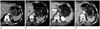
The gastric residual food characteristics and volume were evaluated on the one-year follow-up abdomen-pelvis CT in all the patients by two radiologists (an attending radiologist with 20-year experience of abdominal imaging and a senior radiology resident with 3-year experience). The characteristics of gastric food residue were divided into 5 degrees (0, no residual food; 1, only small amount of radiolucent secretion; 2, poorly defined amorphous food materials; 3, well-delineated measurable food materials; 4, bezoar-like solid food materials) (Fig. 1). The amount of residual food was calculated by the following ellipsoid volume formula for the patients with grade 3 and 4 stasis,
4/3 × π × abc
a = the longest transverse dimension on axial image,
b = the longest anterior-to-posterior dimension on axial image,
c = the longest cranial-to-caudal dimension on coronal image
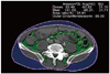
Pre- and post-operative visceral (VF), subcutaneous (SF) and total fat (TF) components were evaluated preoperatively and at one-year follow-up abdomen cross-sectional CT, respectively. All fat components were measured at the umbilicus level using the Aquarius workstation (TeraRecon, Inc., San Mateo, CA, USA) (Fig. 2). The fat tissue area was measured by a standard adipose tissue highlighting techniques, with a fixed attenuation range from -190 to -30 HU, as defined by Sjöström et al. (9). The post-operative fat change was calculated by subtracting the post-operative fat components from the pre-operative fat components. All fat components measurements were performed by one radiologist (a senior radiology resident with a 3-year experience).
Statistical Analysis
All statistical analyses were performed using SAS version 9 (SAS Institute, Cary, NC, USA). We used the t-test, chi square and Fisher's exact tests to analyze the comparison in clinicopathological characteristics between the LSTG and OSTG groups. The interobserver agreement for the characteristics of gastric food residue was evaluated by the kappa value and interobserver reproducibility for the gastric food residue volume measurement was evaluated by the intraclass correlation coefficient and paired t-test. For further statistical analyses, all the discrepant data between 2 observers was unified after repeated review of the CT images in conference. The predictable factors associated with the characteristics of gastric food residue were investigated between the no or negligible gastric stasis groups (degree 0 and 1) and the volume-measurable gastric stasis groups (degree 3 and 4) using logistic regression analysis. The intermediate group showing small amount of amorphous food stasis (degree 2) was not included because of the possibility of temporary finding of food residue, which might be dependent on the fasting time and water ingestion before the imaging and unmeasurability of the scattered food materials in the water-filled stomach. The volume was investigated by the univariate and multivariate regression analyses only for the volume-measurable gastric stasis groups (degree 3 and 4). For the evaluation of gastric food residue characteristics and volume, the patient's age, sex, operation approach (LSTG or OSTG), pT and pN stages, and reconstruction methods (Billroth I or II) were evaluated as the explanatory variables. The post-operative abdominal SF, VF and TF changes between the LSTG and OSTG groups were investigated by the univariate and multiple regression analysis. The preoperative fat was adjusted for this analysis. The predictable factors associated with the postoperative fat change included the above-mentioned factors. Finally, the post-operative fat changes were correlated with the residual food volume by the Pearson's correlation test. For all statistical analyses, the significance was set at p < 0.05.
RESULTS
The comparison of the clinicopathological characteristics between the LSTG and OSTG groups are shown in Table 1. No significant differences were observed between the two groups with respect to age, sex and reconstruction method. However, pT and pN stage were statistically different between the 2 groups (p < 0.001), and the LSTG group contained much more patients with lower pT and pN stages compared with those of the OSTG group. The weighted kappa value of interobserver agreement for the gastric food residue characteristics was 0.91, which means substantial agreement. Intraclass correlation coefficient for gastric food residue volume was 0.995 and the p-value of paired t-test was 0.130, which suggests good interobserver reproducibility.
The LSTG group showed higher frequency of residual food regardless of the degree of stasis (Fig. 3). Although the influence of the operation approach on the gastric residual food characters was not statistically significant (p = 0.072) on the logistic regression analysis (Table 2), LSTG caused higher incidence of the measurable or bezoar-like solid food stasis (degree 3 or 4) compared with OSTG (21.5% versus 12.7%). Meanwhile, age, sex, and pT and pN stages were not all statistically significant factors influencing the gastric food residue characters (Table 2). All the patients with Billroth II showed degree 0, 1, or 2 gastric food residue characteristics with no degree 3 or 4; therefore, the logistic regression analysis for the reconstruction method could not be performed.
The gastric food residue volume for measurable cases (degree 3 and 4 characteristics) was larger in the LSTG group than in the OSTG group (13779 cc ± 14457 in LSTG vs. 6295 cc ± 7027 in OSTG). On the univariate and multiple regression analyses for the predictable factors associated with the gastric food residue volume only for the patients with the degree 3 and 4 characteristics, the laparoscopic approach tended to cause larger volume of residual food, although the influence of the operation approach on the gastric residual food characteristics was not statistically significant (p = 0.137 on univariate analysis and p = 0.059 on multivariate analysis) (Table 3). Due to the absence of the measurable gastric food characteristics (degree 3 or 4) for the patients with Billroth II, the influence of reconstruction method on the gastric food residue volume could not be evaluated.
The average postoperative abdominal fat change was 42.0 cm2 ± 38.8, 28.9 cm2 ± 41.2 and 70.9 cm2 ± 67.0 for VF, SF and TF, respectively. The VF decrease was statistically larger in the LSTG group than in the OSTG group (p < 0.05, β= -7.906) on the univariate analysis (Table 4). However, on the multiple regression analysis with the pT and the reconstruction method as explanatory variables, the VF change was not statically different between the LSTG and OSTG groups (Table 4). The operational approach was not an independent factor that affected the postoperative SF and TF changes on the univariate and multiple regression analyses (p < 0.05). Meanwhile, the preoperative fat and the reconstruction method affected the postoperative VF, SF, and TF changes on the univariate and multivariate analyses (p < 0.05) (Table 4). The patients with Billroth II reconstruction showed more postoperative VF, SF, and TF decreased (p < 0.001, β = -14.708 in VF change, -25.712 in SF change and -41.963 in TF change). All the postoperative VF, SF and TF changes were not correlated with the gastric food residue volume (p values = 0.352 on VF change, 0.815 on SF change and 0.368 on TF change).
DISCUSSION
As a post-operative complication, there have been several endoscopy studies of gastric food stasis after subtotal gastrectomy; Watanabe et al. (2) reported that gastric food residue was observed in 18.7% of a total 374 endoscopic evaluation after subtotal gastrectomy. Jung et al. (1) reported that the incidence of food stasis was 55.5%, 31.9% and 20.9% at 3, 12, and 24 months, respectively on the endoscopic evaluation after subtotal gastrectomy. Depending on the various reconstruction methods, there were some reports about the differences in degrees of gastric food stasis (1, 7). Kubo et al. (7) showed the incidence of residual food in 14.0% of the Roux-en-Y reconstruction group, 22.3% of the Billroth 1 group, and 37.5% of pylorus-preserving gastrectomy group. In addition, Jung et al. (1) mentioned a higher food residue score in the Billroth I group compared with that of the Billroth II group at 3 months after surgery, but there was no difference at 12 and at 24 months. However, in the present study, Billroth I reconstruction showed more gastric stasis compared with Billroth II on one-year follow-up MDCT. We could assume that the gastrojejunostomy at the dependent portion of the remnant stomach encourages food passage after Billroth II reconstruction regardless of the remnant stomach volume. It was mentioned by Behrns and Sarr (10) that post-operative gastric emptying might be more dependent on the restored gastrointestinal continuity rather than on the extent of gastric resection itself.
LSTG have been widely acclaimed as a feasible procedure for gastric cancer in the aspect of technical safety and oncological efficacy (11-16). In spite of the merits of LSTG, our study results showed that LSTG caused higher incidence of postoperative gastric food stasis than OSTG on one-year follow-up MDCT in more severe grades, as well as in the volume of the food residues. According to the previous reports, postoperative food stasis may arises from hypoperistalsis and low gastric acidity after vagotomy and loss of normal pyloric function (5, 10). In terms of more gastric stasis with laparascopic approach in our study, we speculated that there could be a tendency toward larger volume of the remnant stomach with LSTG, because of a lower grade tumor stage compared with the open abdominal surgery; although, we did not have any residual volume data of the remnant stomach after the partial gastrectomy in the present study. We could assume that the larger gastric volume after LSTG could facilitate more gastric stasis especially in Billroth I gastroduodenostomy, which was not reconstructed along the gravity-dependent direction of food passage like Billroth II gastrojejunostomy. In a previous report by Jung et al. (1) the gastric food stasis was not related with the remnant gastric volume measured by the ratio of lesser curvature length in the remnant stomach to that of the entire stomach. However, the measurement of actual gastric volume is difficult in the most basic sense, either by endoscopy or MDCT, because of the elasticity of gastric wall and unpredictable gastric movement. In addition to the volume of the remnant stomach, there could be a possibility of luminal distortion around the anastomotic site by LSTG. Without the bare finger sensation and naked eye view during the laparoscopic surgery, the tension and the direction of resection margin between the stomach and the duodenum could be unevenly matched to each other during the procedure of anastomosis compared with the open surgery. In such cases, there may be a higher possibility of functional obstruction at the anastomotic site.
In the present study, we investigated the influence of gastric food stasis on the abdominal VF, SF and TF by MDCT. Weight loss is one of the common problems after gastrectomy, and the main mechanisms are implicated in poor oral uptake and impaired absorption (17, 18). Several investigators reported that weight loss occurred primarily during the early postoperative period (6 months to one year) and was mainly caused by loss of body fat (19-22). In an investigation by Yoon et al. (23) patients who underwent gastrectomy lost TF, VF and SF during the postoperative period up to six months and the decrease in VF was greater and more persistent than the decrease in SF on CT. It would be due to more distinct metabolic characteristics of VF that visceral adipocytes are metabolically active and respond more promptly to metabolic stimuli (24). Similar to the previous study (23), VF was decreased more than SF after one year of gastric surgery in the present study. In the aspect of the relationship between the abdominal fat change and the way of surgical approach (LSTG versus OSTG), our results showed that only VF was more decreased in the patients with LSTG compared to those with OSTG. When we consider the pT on the multivariate analysis, the different operational approach was not a statistically independent factor affecting the postoperative VF change. Furthermore, the VF, SF, and TF changes were not correlated with the gastric food stasis volume. Similar to a previous study showing no significant association between the food retention and the body weight changes (1), our results suggest that the degree of postoperative gastric food stasis may not have much of an affect the metabolic body fat loss. Postoperative abdominal fat change appears to be multifactorial.
There were several potential limitations of our study. First of all, although there was a tendency that LSTG caused more postoperative gastric food stasis, we could get just marginal statistical difference in both characteristics and volume of gastric food residue between both groups, which might be related to the limited size of the study population. In an additional statistical calculation, the required population size was 553 and 112 patients (80% power at a 0.05 significance level) to acquire the statistical difference for the gastric food characteristics and volume between both groups. Second, the clinicopathological characteristics between both groups were not all equal to each other; the OSTG group included the patients with higher stages in pT and pN. To overcome this limitation, we tried multiple regression analysis by evaluating the multiple explanatory variables including the pT and pN stages. In terms of body fat changes, however, the patients with higher cancer stage could have already experienced more fat loss before the first CT examination prior to the surgery already. In such circumstances, the implication of direct comparison of body fat between the pre- and post-operation CT can be limited. Third, because of the nature of our study, the clinical aspect of the gastric food stasis, such as degree of symptoms or quality of life, was not evaluated. Although the postoperative gastric food retention hardly affect the abdominal fat change and it's clinical significance in terms of symptoms and quality of life is not elucidated in our study, we believe that any effort to reduce the gastric stasis has to be continued for proper endoscopic field of vision and to minimize the carcinogenic effect from gastric food residue itself. For example, an alternative reconstruction method may be required for subtotal gastrectomy and gastroduodenostomy, especially for the laparoscopic approach, like the anastomosis at the posterior wall of greater curvature side of the stomach. Continuous and thorough diet education could be essential for patients undergoing gastric cancer surgery, especially for those with LSTG. Fourth, the degree of food stasis was determined depending on the subjective findings of MDCT. With the reference of the endoscopic grading of the residual food, which the National Cancer Center had arbitrarily determined by referring to the classification system reported by Kubo et al. (7), we classified the characteristics of food stasis into 5 degrees considering the attenuation densities of solid components distinguished from the ingested water before the imaging and the contour of the gastric residues on MDCT. Meanwhile, we could get substantial interobserver agreement (the weighted kappa value, 0.91), which meant a high degree of reliability for our classification of food stasis in the present study. Finally, although the gastric food stasis volume could be related to the remnant gastric volume as a possible explanation for larger gastric stasis in the LSTG group, we were not able to investigate the remnant gastric volume on CT because of the fact that the stomach is not a fixed solid organ, and further investigations would be needed for the gastric volume measurement.
In conclusion, compared with OSTG, LSTG tends to develop gastric stasis more frequently without discernable differences in postoperative body fat changes between the 2 different surgical approaches. Although the implication of a higher incidence of gastric food stasis in the patients after LSTG seems not so obvious in the present study, any effort to reduce the food stasis after subtotal gastrectomy, especially LSTG, should not be neglected to reduce the pre-established problems that include a limited field of view for endoscopy and the carcinogenic potential.


 XML Download