

 PDF
PDF ePub
ePub Citation
 Print
Print


INTRODUCTION
Juvenile xanthogranuloma (JXG) is a proliferative histiocytic disorder experienced during childhood and adolescents, and most commonly presents in the first two decades of life (1). JXG is not a true neoplasm, but rather a reactive proliferation of histiocytes, and belongs to the category of non-Langerhans dendritic cell disorders. However, the etiology and pathogenesis of JXG are still unknown. Most cases present as a solitary cutaneous lesion followed by a soft tissue mass in the head and neck region. Extracutaneous JXG, especially when involving only the spinal column, is extremely rare in adults. Through a review of the literature, we identified only three previous cases, which were in a 38-year-old male, 29-year-old male, and 41-year-old female (2-4). We describe a 67-year-old female who presented with an intradural-extramedullary (IDEM) tumor of the spinal cord. To our knowledge, this case report represents the oldest patient with a new presentation of spinal JXG. We report magnetic resonance imaging (MRI) findings of JXG of the spinal cord, which were then pathologically confirmed.
CASE REPORT
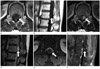
A 67-year-old female presented with a complaint of lower back pain with a tingling sensation in the left leg running from her left thigh to left calf of two months in duration. She had no other complaints. Her only significant past medical history was controlled hypertension. General physical and neurologic examination did not reveal any abnormalities apart from slight hyperactivity of the deep tendon reflex in both knee jerks. Muscle strength in all limbs, gait, and co-ordination were normal, and there was no bowel or bladder dysfunction. Cranial nerve examination and higher executive functions were normal. Lumbar spinal MRI with contrast enhancement showed an approximately 1.7 × 0.9 cm oval-shaped well-defined IDEM lesion at the L1 level. This lesion exhibited mixed signal intensity on the T1-weighted image (WI) and high signal intensity on T2-WI. Central homogenous enhancement was observed after contrast administration (Fig. 1).
The patient underwent a left hemilaminectomy at L1 and a partial hemilaminectomy at T12 for removal of the tumor. A yellowish soft mass was located in the intradural and extramedullary areas, and a total tumor resection was performed.
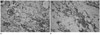
Histologic review demonstrated a collection of mononuclear cells with dilated blood vessels and areas of focal hemorrhage. The mononuclear cells were moderate to large round cells with small and uniform nuclei and lipid-laden cytoplasm (Fig. 2A). The cells stained positively for CD68, a histiocyte marker (Fig. 2B). The pathologic diagnosis was JXG.
Postoperatively the patient's pain and tingling sensation were relieved. A postoperative MRI obtained at 1 and 3 years after surgery did not reveal any residual or recurrent tumor.
DISCUSSION
JXG is the most common form of non-Langerhans cell histiocytosis. The exact etiology or pathogenesis of JXG has not been determined, although it is regarded as a reactive process rather than as a neoplasm. The incidence of JXG has not been estimated, but may be higher than is generally recognized as JXGs occur early in life and usually resolve spontaneously. Based on an autopsy series, Ayres and Haymaker (5) suggested that the incidence varies from 1.6% to 7% of the general population. Despite the use of the term "juvenile", development of this disease during adulthood is possible, and generally occurs in the late 20s to early 30s (6). Predominance in males has been reported in childhood cases, but there is no difference by sex in adults. JXG typically presents as cutaneous lesions. Extracutaneous involvement is not common, with an incidence of only 5%. The eye is the most common site of extracutaneous involvement, and other involved organs include the oropharynx, heart, lung, liver, spleen, adrenal glands, muscles, subcutaneous tissues, and the central nervous system (7, 8). Solitary JXG involving the spinal column is extremely rare in adult patients. Thus far, only three cases have been reported in the English-language literature. Two of the reported cases involved the cervical spine and one was a lumbosacral lesion. The symptoms in all cases were a result of cord compression by the lesion (2-4).
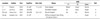
MRI has been demonstrated to be the best imaging modality for the localization of spinal tumors and evaluation of their correlation with adjacent structures. The differential diagnosis contains some IDEM tumors, including spinal ependymoma, astrocytoma, and schwannoma. Spinal ependymoma and astrocytoma can be considered in the location of the lesion. Ependymoma is a tumor that arises from the ependyma, a tissue of the central nervous system. Generally, in pediatric cases the location is intracranial, while in adults it is spinal. Their occurrence seems to peak at age 5 years and then again at age 35. Ependymomas are generally hyper-intense on T2-WI and hypo-intense on T1-WI, which is often heterogeneous. Ependymomas nearly always show contrast enhancement, though are not always homogenous (9). Astrocytomas are the most common glioma and can occur in most parts of the brain and occasionally in the spinal cord. People can develop astrocytomas at any age. Astrocytomas are iso- to slightly hypo-intense on T1-WI, hyper-intense on T2-WI, and commonly have associated cysts. They enhance less intensely and are more eccentric than ependymomas (10). Schwannomas can be considered in the shape of the lesion. Schwannomas frequently affect the eighth cranial nerve most commonly. Less commonly, schwannomas occur in other nerves that contain Schwann cells. These tumors are most common among people who are 50 to 60 years of age. Schwannomas are generally hypo or iso-intense on T1-WI and hyper-intense on T2-WI which is often heterogeneous, and are frequently associated with hemorrhage, intrinsic vascular changes, cyst formation, and fatty degeneration. The location of the spinal JXG is variable from the cervical spine to the sacrum. The age spectrum of the adult spinal JXG is also wide from twenty nine to sixty seven. Spinal JXG may appear with variable signal intensity, that is, a mixture of hypo-, iso-, and hyper-intense in T1-WI and T2-WI. Furthermore, the lesion may exhibit homogenous enhancement after contrast media administration (Table 1).
Upon gross anatomic examination, the JXG is a well-encapsulated and round lesion with a yellowish surface. JXG is confirmed by the microscopic pathologic identification of foamy histiocytic cells and an immunohistochemical finding of mononuclear cells, giant cells, and spindle cells positive for lysozyme stain and CD68 but negative for CD1a and S-100 proteins, which are all reactive markers of Langerhans cells (Fig. 2B).
These tumors may grow slowly without regression, and thus the symptoms worsen gradually. This is the distinguishing and important characteristic of JXG in adult patients compared with that found during childhood and adolescent JXG, which regresses spontaneous. Despite being a pathologically benign tumor, it is conventional to remove as much of the tumor as possible. There have been no reports of recurrence after total mass excision (4).
We have described the case of a 67-year-old female with a solitary tumor identified as a JXG involving the spinal column. Although JXG is very rare, JXG should be considered in the differential diagnosis of IDEM tumors of the spinal cord in adult patients.
 XML Download