

 PDF
PDF ePub
ePub Citation
 Print
Print


INTRODUCTION
Although many women participate in breast screening programs, multidetector computed tomography (MDCT) often provides the first images of the breast when patients undergo chest MDCT for evaluation of pulmonary or cardiac disease (1-4). Since an MDCT scan provides improved contrast resolution, a larger field of view, and has cross sectional capability, breast lesions may be viewed more easily with a MDCT scan (1, 5, 6). With the increasing use of MDCT, a growing number of unexpected breast abnormalities of uncertain clinical significance have been observed. In practice, few studies have demonstrated the significance of incidentally detected breast lesions on MDCT (3, 4). The purpose of this study was to evaluate the significance of incidentally detected breast lesions during assessment of chest related symptoms on a chest CT scan.
MATERIALS AND METHODS
Patients
Our study was approved by the institutional review board and the informed consent requirement was waived because of the retrospective design of the study. Between March 2007 and March 2010, 25398 female patients who exhibited breast lesions on a sonochest CT scan were found in our institutional database. Among the 3745 patients who had breast lesions found on a chest CT scan, women with known breast cancer and post operative follow up for breast cancer were excluded. Among fifty-four patients who were incidentally identified as having abnormal breast lesion on a chest CT scan, 26 patients had breast sonography performed within 3 months according to our institutional data-base. Among thirty-six lesions for 26 patients, six patients were excluded due to being unavailable for a follow-up study or pathological confirmation of lesions. Finally, a total of twenty-four breast lesions in twenty patients were reviewed and chest CT findings were compared with sonographic findings retrospectively (Table 1). All patients underwent chest CT for the evaluation of chest abnormalities such as an abnormality on a plain radiography (n = 2), chronic cough (n = 1), hemoptysis (n = 1), bronchiectasis (n = 1), asthma (n = 2), chest pain (n = 3), palpitation (n = 1), traumatic contusion (n = 2), work up of lung cancer (n = 2), hepatoma (n = 1), and fever of unknown origin (n = 1), chest wall mass (n = 1), post operative work up of colon cancer (n = 1), and soft tissue sarcoma (n = 1). The median age of our study population was 50 years (range: 26-78 years).
Imaging Technique
Chest CT was performed in the supine position using one of the six MDCT machines: Siemens 4-slice, 16-slice, 64-slice CT system (Siemens Medical System, Forchheim, Germany); Light-Speed VCTXe (GE Healthcare, Milwaukee, WI, USA); GE LightSpeed Ultra 8 slice, 64-slice Lightspeed VCT (GE Healthcare, Milwaukee, WI, USA). The chest CT was performed with the following parameters: 120 mAs, 120 kVp, 3 mm thickness, 64 × 0.6 mm detector collimation; 100 mAs, 120 kVp, 5 mm thickness, 16 × 0.75 mm; 120 mAs, 120 kVp, 5 mm thickness, 64 × 0.625 mm; 340 mAs, 120 kVp, 5 mm thickness, 16 × 1.25 mm, 170 mAs, 120 kVp, 5 mm slice, 64 × 0.625 mm. One breath-hold acquisition was obtained 55 seconds after an IV rapid bolus administration of nonionic contrast material. All patients underwent MDCT in the supine position. Multiplanar reconstructions (axial and coronal) were used for the evaluation of breast lesions. The images were obtained by using a standard soft-tissue algorithm [window width, 480 Hounsfield units (HU); level, 34 HU]. One patient underwent a contrast-enhanced scan and 19 patients underwent both unenhanced and contrast-enhanced scans on one of the six MDCT machines. Sonography was performed by a physician using a broad-band-width linear-array transducer with a center frequency of 10 MHz supplemented by a transducer with a center frequency of 7.5 MHz (GE LogiQ 700 Expert Series; GE Medical Systems, Milwaukee, WI, USA; HDI 5000; Advanced Technology Laboratories, Bothell, WA, USA; IU-22, Philips, Bothell, WA, USA). Breast ultrasonography was performed in a mean time of 19 days (range, 1 day-3 months) following the day on which breast lesions were detected on the chest CT scan and sonographic findings were correlated with CT findings.
Image Interpretation
Twenty-four chest CT detected lesions correlated with sonographic findings. Breast density on CT was divided into four patterns using the Breast Imaging Reporting and Data System (BI-RADS) classification for mammographic parenchymal density: pattern 1, a fatty breast; pattern 2, a fatty breast with scattered fibroglandular densities; pattern 3, a heterogeneously dense breast; pattern 4, an extremely dense breast. CT features were assessed in consensus by two radiologists who were blinded to breast imaging or pathology results. Incidentally detected breast masses on chest CT scans were analyzed for size, location, shape (oval, round, irregular), margin (circumscribed, non-circumscribed), enhancement pattern (homogeneous, heterogeneous) and the level of HU. Sonographic results were categorized using the BI-RADS lexicon according to shape (oval, round, irregular), margin (circumscribed, non-circumscribed) and echogenicity (isoechogenicity, hypoechogenicity).
Statistical Analysis
The statistical analysis was performed using the chi-squared test and Mann-Whitney test. The Fisher's exact test was used for analysis of the correlation between sonographic and CT findings. A p value of 0.05 was chosen as the threshold for statistical significance. SPSS software was used (SPSS version 14.0, SPSS, Chicago, IL, USA).
RESULTS
Pathologic Outcomes
The pathological diagnosis was confirmed in twenty-two lesions. Biopsy procedures performed included an ultrasound guided 14 G core needle biopsy (n = 15), mammotome biopsy (n = 1), and excisional biopsy (n = 6). Pathologic findings included infiltrating ductal carcinoma (n = 7), benign lesions [n = 15; fibroadenoma (n = 10), and fibrocystic disease (n = 5)]. Two patients underwent follow-up sonography after 20 months and 24 months, respectively. In one patient a well circumscribed, round, inhomogeneous enhancing mass was detected on CT scan with sonographic correlation and no significant change in the mass was observed on a follow-up sonography performed after 24 months. In the other one patient, an irregular, microlobulated, inhomogeneous enhancing mass was detected on CT scan with negative findings on sonography. In this patient, no interval change was seen in the detected abnormal mass on a follow-up sonography after 20 month. The mean size of incidentally detected lesions on chest CT scans was 1.6 cm (range: 0.5-6.3 cm).
Image Analysis of Incidentally Detected Breast Lesions
Among 36 lesions of 26 patients that were incidentally detected breast lesions on a chest CT scan, 31 lesions were correlated with sonography in 86% (31/36). Exclusion of 12 lesions was due to subjects being unavailable for pathologic results or follow up sonography, seven (29.2%) lesions were malignant and 17 (70.8%) lesions were benign lesions.
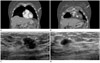
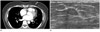
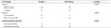
Estimation of breast density on CT using the BI-RADS classification for mammographic parenchymal density was pattern 1 (n = 0), pattern 2 (n = 6), pattern 3 (n = 8), and pattern 4 (n = 6). When evaluation of the twenty-four masses on CT scan was performed, the shapes of the lesions were oval (n = 2), round (n = 10), and irregular (n = 12). The margins were circumscribed (n = 10), and noncircumscribed (n = 14). The contrast enhancement patterns were homogeneous (n = 5) and heterogeneous (n = 19). The mean measured HU of the mass was 39.6 (range: 14-47 HU) on pre-enhanced CT scan and 113.79 HU (range: 50-236 HU) on contrast-enhanced CT scan (Table 2). An analysis of CT findings for incidentally detected lesions revealed a significant difference between benign (n = 17) and malignant (n = 7) lesions in terms of shape and margin (p = 0.007; p = 0.008, respectively) (Figs. 1, 2); and there was no significant difference in benign and malignant lesions between pre-enhanced CT scans and contrast enhanced CT scans (p = 0.107). No significant difference was observed in HU between benign (mean HU; 105.71) and malignant lesions (mean HU; 133.43) (p = 0.120). A total of 22 lesions were pathologically confirmed by sonographic BI-RADS classification as category 3 (n = 10), category 4 (n = 8) and category 5 (n = 4). An analysis of correlation between characteristics of CT findings and sonographic findings for shape, margin and enhancement (echogenicity) demonstrated a significant correlation in terms of shape and margin (p = 0.001, respectively) (Table 3).
DISCUSSION
The majority of breast lesions are diagnosed through the breast screening programs and clinical breast examination. However, with the increasing use of MDCT for detection of other pathologies, new breast lesions are being increasingly detected incidentally during CT scans for diseases other than those of the breast (7, 8). CT scans are not only sensitive for detection of small breast lesions within dense breasts but also allow for better visualization of some breast lesions adjacent to the chest wall (4, 7, 9). Usually, chest CT scans are reported by radiologists who do not specialize in breast imaging and pathology. Therefore, these breast lesions may be missed or may not be mentioned in radiological reports nor would such cases be referred to the breast specialist for assessment and management.
Several CT techniques have been used in assessment of breast lesions including contrast-enhanced CT scans, multi-detector techniques MDCT, and dual time positron emission tomography-computed tomography (7). For an accurate assessment of breast lesions, standardized terminology is required for description and categorization of abnormal findings using the BI-RADS lexicon of the American College of Radiology (1, 5). The features of breast lesions may be suggestive of benign or malignant breast pathology (4-7). The CT predictive features of breast malignancy include an irregular or spiculated margin, irregular shape, rim enhancement, and marked early and/or peripheral enhancement (1, 4, 5). Indeterminate breast lesions or masses showing benign features on CT also require additional validation of benignity using diagnostic mammography with or without sonographic evaluation (1). In our study 29.2% (7/24) of incidental breast lesions were proven to be malignant on CT scan. According to several reports, 24-32% of incidental breast lesions were subsequently proven to be malignant (3, 7, 8). Lin et al. (4) reported that an irregular margin of incidental enhancing breast lesions can be suggestive of malignancy and a much higher rate (70%) of malignancy was demonstrated in lesions detected incidentally on CT scans. In our study, an irregular shape or a non-circumscribed margin was shown to be a statistically significant factor for differentiation between benign and malignant breast lesions. Randomized blinded multicenter trials with larger sample sizes are needed for the characterization of the sensitivity and specificity of contrast enhanced breast CT.
Prionas et al. (10) demonstrated that the use of a contrast-enhanced breast CT could deliver significantly improved conspicuity of malignant breast lesions, including ductal carcinoma in situ. They also demonstrated that malignant breast lesions show significantly greater enhancement with higher HU counts than do benign lesions. In our study, there was no significance regarding HU of breast lesions between benign and malignant breast lesions. In our study, chest CT was performed for the evaluation of chest lesions. Therefore, enhancement was performed 55 seconds after contrast injection without dynamic enhancement. If the CT scan was performed with a focus on the breast lesion, quantification of lesion enhancement during contrast-enhanced breast CT could potentially aid in the prediction of malignancy. Porter et al. (3) reported that 50% of normal breast glandular tissues mimic pseudo-masses that may not be enhanced on contrast-enhanced CT.
This study has several limitations. First, this study was designed retrospectively. Therefore, small numbers of selected patients having incidental breast lesions on a chest CT scan with breast sonography were included in this study. In addition, there is a possibility that a few patients whom breast lesions have not been reported on chest CT readings may not be included. In our cases, all of the lesions were mass lesions and non-mass lesions or calcifications were not included. That is because the study was designed retrospectively with selected patients having reported breast lesions on chest CT scans. Second, the same protocol was not used for the performance of CT examinations. With the increasing use of MDCT, a growing number of unexpected breast abnormalities have been observed and CT scans could detect small breast lesions that were not detected during the screening program. Therefore, the use of MDCT is advocated for breast evaluation when incidental enhancing breast lesions are detected on a routine chest CT scan.
In conclusion, the radiologist should evaluate the breasts on chest CT performed for other thoracic indications. If a breast lesion is detected incidentally on a chest CT scan, additional breast examinations should be performed. An irregular shape or a non-circumscribed margin of breast lesions can be considered as a suggestive sign of malignancy.
 XML Download