

 PDF
PDF ePub
ePub Citation
 Print
Print


INTRODUCTION
Several different injectable materials have been used for breast augmentation in females, such as liquid silicone, autologous fat, liquid paraffin, and polyacrylamide gel (1-3). Imaging findings for each material are well-known, with typical mammographic and sonographic features; hence, their presence is readily diagnosed. However, a spiculated mass or diffuse infiltrative change can sometimes mimic breast malignancy or other systemic diseases (4). In this report, we present a bilateral diffuse infiltrative disease that mimics the imaging findings of a male patient who underwent foreign material injection for breast augmentation.
CASE REPORT
A 64-year-old male patient was presented to our breast center with a diagnosis of gradual breast enlargement when he was an inpatient at our hospital for cerebral infarction. On physical examination, the patient had bilateral enlarged breasts, retracted nipples, and scarring on the mid-upper side of the left breast.
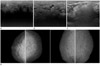
An ultrasound examination was initially undertaken for gynecomastia. A two-dimensional ultrasound (IU22, Philips Medical System, Bothell, WA, USA) with a high-frequency probe (5-12 MHz linear transducer) showed bilateral, severely thickened skin and diffuse edematous change along the subcutaneous fat layer. A few cysts, which were variable in size and round-to-oval hypoechoic masses, were seen in both breasts. In the center portion of both breasts, there were ill-defined hypoechoic lesions with markedly posterior acoustic shadowing. Also, large hyperechoic mass-like lesions were noted in both axillae (Fig. 1A-C). A standard two-view mammography was performed, which demonstrated marked enlargement of both breasts with diffusely increased density, diffuse surface skin thickening, and trabecular thickening. Furthermore, the nipple of the left breast was retracted (Fig. 1D).
First, we suspected diffuse infiltrative disease, such as lymphoma or diabetic mastopathy. We did not consider the possibility of interstitial injection, because the patient was a normal married man. Accordingly, we recommended an ultrasound-guided biopsy; however, he did not want to undergo further examination and ultimately, left the hospital.
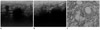
About 14 months later, the patient revisited our breast center due to exacerbated breast enlargement and heating sensation. He underwent an ultrasound examination again as well as an ultrasound-guided core biopsy using a 14-gauge needle, which led to the pathological diagnosis of lipogranulomatous inflammation with severe sclerosis in both breasts (Fig. 2). A pathologist recommended that we should check for a history of interstitial injection. The patient then admitted that he had undergone bilateral breast injection with his friends just for fun almost 30 years ago. He was not forthcoming about the details of this procedure.
DISCUSSION
The injection of various materials for breast augmentation was first used in the early 1900s (2, 3). Several injectable materials, such as liquid silicone, autologous fat, liquid paraffin, and polyacrylamide gel have been used for breast augmentation in females. Liquid silicone injection appears on mammography as well-defined, rounded, peripherally calcified masses (2). Liquid paraffin injection appears as circumscribed, noncalcified masses, streaky opacities, architectural distortion, or dystrophic or ring-like, calcified indistinct mass (paraffinoma) on mammography (2, 3). Autologous fat injection is presented as various forms of fat necrosis on mammography. Polyacrylamide gel injection appears as a single or multiple fluid collections in the retroglandular area on mammography (1, 5). These relatively typical findings of foreign material injection for breast augmentation have been reported mostly in female patients.
In this report, we describe the imaging findings of foreign material injection (paraffin) for breast augmentation in a male patient. To our knowledge, this is the first case report of foreign material injection for breast augmentation in a male patient. Our patient complained of gradual breast enlargement. Mammography demonstrated markedly enlarged breasts with increased density, combined with skin and trabecular thickening. An ultrasonography demonstrated a few oil cysts and ill-defined hypoechoic lesions with posterior acoustic shadowing in both breasts. Enlarged lymph nodes with central echogenicity were noted in both axillae. Thus, diffuse infiltrative disease, such as lymphoma or diabetic mastopathy, was strongly suspected. There are two types of lymphoma of the breast: the solitary or multiple mass-forming type and the infiltrative type (6). In the infiltrative type of lymphoma, diffuse opacification and trabecular and skin thickening may be present on mammography, whereas irregular oval hypoechoic masses with posterior acoustic shadowing may be present on sonography. Unilateral or bilateral abnormal axillary nodes are the most common mammographic finding in systemic lymphoma. Diabetic mastopathy occurs primarily in patients with a history of long-term insulin-dependent (type 1) diabetes. Mammography demonstrates asymmetric density with ill-defined margins; however, it does not show microcalcifications or dense glandular tissue. Sonography shows a hypoechoic mass or marked acoustic shadowing (7).
Pathological examination revealed lipogranulomatous inflammation with severe sclerosis, compatible with the injection of paraffin. Lipogranulomatous or oleogranulomatous mastitis is a well-known complication following the injection of melted petroleum jelly, such as liquid paraffin or silicon, into the breasts (8). There are two presenting types of oleogranulomatous mastitis: suppurative and nonsuppurative. In the suppurative type, masses may ulcerate and become infected to form a discharging sinus or a fistula tract. The surface skin of the breast may also shows a brownish discoloration. An inverted nipple, peau d' orange or enlarged lymph node may be present. However, unlike suppurative mastitis, the skin overlying the breast is often normal in the nonsuppurative type (9).
In conclusion, foreign material injection for breast augmentation in males shows various imaging findings. Sometimes these are strongly suggestive of malignancy or other systemic disease. Radiologists should be familiar with the spectrum of the appearance of foreign material injection for breast augmentation. Regardless of sex, detailed history taking and pathological confirmation by means of core-needle biopsy are important for the prevention of misinterpretation and, by extension, mistaken treatment.
 XML Download