

 PDF
PDF ePub
ePub Citation
 Print
Print


INTRODUCTION
Inflammatory myofibroblastic tumor (IMT) is a rare mesenchymal neoplasm that is typified by a myofibroblastic and mixed inflammatory cell infiltrate (1). The tumor presents itself most commonly in children and young adults, and it occurs most frequently in the lungs (1). Although IMT has been reported in various extrapulmonary sites, its occurrence in the ileum is uncommon. Furthermore, its presentation as intussusceptions is rare and thus, only a few cases have been reported (2). To the best of our knowledge, there is no report in the radiology literature regarding IMT-induced intussusception.
We report multidetector computed tomography (CT) imaging findings of an IMT of the ileum in an adult causing an ileoileal intussusception, and further discuss its radiological and pathological features.
CASE REPORT
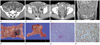
A 55-year-old man was admitted to our hospital with a one-week history of abdominal pain. He had no history of any surgical operation and systemic disease. A physical examination revealed a distended abdomen with tenderness in the entire abdomen as well as hyperactive bowel sounds. Plain abdominal radiographs revealed dilated small bowel loops with different heights of air-fluid level and a thickening of valvulae conniventes with mechanical ileus. Contrast-enhanced abdominal CT demonstrated dilated small bowel loops and invagination of a segment of the distal ileum with mesentery into the adjacent thick-walled intussuscipiens (Fig. 1A, B). A 2.5 cm sized lead mass with enhancing thin wall in the distal portion of the intussusceptum was identified. The CT attenuation value of the mass was measured at about 34 Hounsfield units (HU) on the unenhanced image and approximately 41 HU on the enhanced image (Fig. 1C, D). There was a small amount of pelvic ascites and no abnormal lymphadenopathy. The differential diagnosis included Meckel diverticulum, duplication cyst, gastrointestinal stromal tumor, or leiomyoma with intussusception.
An exploratory laparotomy revealed ileoileal intussusceptions at the distal ileum along with edematous small bowel proximal to the lesion. The patient underwent a segmental resection of the intussuscepted ileum as well as an ileoileostomy.
Grossly, the resected specimen of the ileum measured 8 cm in length and 3.5 cm in circumference. There was a solid, well-defined, and intraluminal pedunculated mass measuring 2.5 × 2.0 cm in the ileum (Fig. 1E). The cut surface of the mass revealed a whitish tissue and there was no hemorrhage or necrosis in the mass (Fig. 1F). Microscopically, the well-demarcated tumor involved a submucosa and a muscle layer. The tumor was composed of an admixture of proliferating the spindle cells and inflammatory cells embedded in a highly vascularized stroma. There were mild cellular atypia and mitotic figures (4/50 high power field) (Fig. 1G). Immunohistochemistry of the tumor cells was positive for the smooth muscle actin (Fig. 1H). The resected tumor of the ileum was diagnosed as polypoid IMT.
DISCUSSION
IMT is a rare mesenchymal neoplasm that is typified by a myofibroblastic and mixed inflammatory cell infiltrate (1). Since its first description in 1937, the understanding of IMT has evolved from a reactive inflammatory process to a neoplasm of an uncertain malignant potential (2). Disagreement and uncertainty about the histogenesis of IMTs has resulted in a number of synonyms, which include plasma cell granuloma, plasma cell pseudotumor, inflammatory myofibrohistiocytic proliferation, omental-mesenteric myxoid hamartoma, and, most commonly, inflammatory pseudotumor (3).
The etiology of IMT is poorly understood. Since IMT may be seen following abdominal surgery and trauma, it may represent an infectious or inflammatory process (4). However, based on the role of oncogenic viruses and cytogenetic abnormalities, including the anaplastic lymphoma kinase gene rearrangements on chromosome 2p23, clonal chromosome abnormalities, and DNA aneuploidy, a neoplastic origin for IMT has been supported more recently (2, 3).
IMT occurs most commonly in the lungs. Extrapulmonary sites have also been reported, which include the mesentery and omentum, genitourinary tract, gastrointestinal tract, retroperitoneum, pelvis, head and neck, trunk, and extremities (1). Among extrapulmonary IMTs, 43% of the sites arise from mesentery and omentum whereas the small bowel accounts for only 1.2% of the affected sites (1). Moreover, IMTs arising from the small bowel causing intussusception are rare (2, 3). Intussusception is primarily a disease among infants and children, and about 5% of the cases occur in adults. Whereas 90% of childhood-type intussusceptions are idiopathic, an underlying pathologic process is usually identifiable in over 90% of adult cases (5). Small bowel intussusception without a lead point is more common than intussusception with a lead point. A lead point intussusception involving the small bowel is generally due to a benign condition and less often due to a neoplasm, which is usually a metastatic lesion (6).
IMTs arising from small bowel causing intussusception are extremely rare. Although it is hard to discern the exact underlying disease on radiologic examination, as long as IMT can cause an intussusception, the tumor should be considered as one of the causes of intestinal obstruction.
The clinical presentation of the IMT depends to some extent on its site of origin. Patients with intra-abdominal tumors may also be present with abdominal mass, abdominal pain, vomiting, constipation, and bowel obstruction. IMT may be a cause of chronic obstruction and of acute obstruction owing to intussusception (3).
Although the radiological examinations confirm obstructive features and the presence of intra-abdominal mass lesions, IMT is not a radiologic distinct entity (7). There are many factors that affect the appearance of an intussusception. These include the presence of a lead point, the configuration of the lead mass, the degree of bowel wall edema, and the amount of invaginated mesenteric fat (6). Identification of a lead mass that is separate and distinct from bowel loops is not easy. However, a mass that is seen on CT can serve as a reliable radiologic indicator of intussusceptions with a lead point, even though it is hard to discern the exact underlying disease in most cases (6). The CT appearance of IMT is variable. The mass may be hypoattenuated or isoattenuated on unenhanced scans; moreover, calcification has been observed in the cases of the pancreas, stomach, and liver. Enhancement usually occurs but is not pronounced, and a variety of patterns have been noted including early peripheral, with delayed central filling, and heterogeneous, homogeneous, and no enhancement (8). In our case, the CT attenuation value of the mass measured at about 34 HU on the unenhanced image and approximately 41 HU on the enhanced image.
The combination of conventional hematoxylin and eosin staining and appropriate immunohistochemical staining pathological studies can reliably distinguish IMTs with other mesenchymal tumors (9). The confirmation of the anaplastic lymphoma kinase protein/gene rearrangement is also useful in distinguishing IMT from other spindle cell neoplastic mimickers (3).
The mainstay of treatment for this tumor is surgical resection with wide margins. Incompletely resected tumors may have local recurrence within 1 year; however, recurrence has been seen up to 9 years after resection of the primary tumor (10).
In conclusion, ileoileal intussusceptions rarely occur in adults, particularly that due to IMT. It is hard to detect the exact underlying disease of intussusceptions on radiologic examination. Although intra-luminal IMT of small bowel loop may be a rare cause of intussusception, IMT may be considered as one of the causes of intestinal obstruction.
 XML Download