

 PDF
PDF ePub
ePub Citation
 Print
Print


INTRODUCTION
Malignant melanoma accounts for 1.5% of all reported cancers with prevalence increasing in the past decade. Skin is the most common site for primary malignant melanoma, but it can involve virtually every organ system where melanin cells exist. They can frequently metastasize to the liver, lung, brain and bone before the primary lesion in the skin is clinically evident (1). Here, we report a case of malignant melanoma representing superior mediastinal mass without extrathoracic primary lesion.
CASE REPORT
A 32-year-old man was presented with 3 weeks' history of hoarseness and had been suffering from right ptosis for 6 months. His laryngoscopic examination revealed right vocal cord palsy without vocal cord abnormality. He had no definite past medical history, except for vitiligo whenever he gets sunlight. He had a history of resolved vitiligo on both cheeks and left medial malleolar area, which was retrospectively noted.
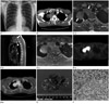
Initial chest radiograph (Fig. 1A) showed a right apical mass-like opacity showing lobulated contour and obtuse angle with adjacent pleura. The patient subsequently had a chest CT scan, which demonstrated a lobulated mass (7.4 × 4.2 × 5.8 cm) with heterogenous enhancement in the right superior mediastinum (Fig. 1B). Tracheal deviation, and obliteration of ipsilateral tracheoesophageal groove by the mass was noted, and this could explain hoarseness with the involvement of ipsilateral recurrent laryngeal nerve. Two days later, magnetic resonance (MR) imaging of the cervical spines was performed on a 1.5-T MR unit. The superior mediastinal mass was mildly hyperintense to muscle on T1 and T2-weighted images (Fig. 1C). The mass contained a small area that was hyperintense on T1-weighted images and hypointense on T2-weighted images. The mass showed invasion of right neural foramen of the 2nd and 3rd thoracic vertebrae and erosion of the adjacent right 2nd rib, which might have caused right side ptosis. Positron emission tomography/computed tomography scan (Fig. 1D) showed high standard uptake values (SUV) uptake (max SUV: 8.25) of the mass and no evidence of metastatic diseases, other than the thoracic lesion. There was no evidence of primary melanoma outside the mediastinum.
The patient underwent total excision of the right superior mediastinal mass (Fig. 1E, F). Gross pathologic examination showed an irregular dark brownish mass measuring 8.3 × 4 × 4 cm and weighing 54 g. Serial sections revealed multiple lobulated tan-brown areas of soft to firm tissue with areas of hemorrhage and necrotic foci. Histologic analysis of the mass revealed large pleomorphic cells with prominent nucleoli, some of which contain melanin pigments. A Fontana-Masson stain confirmed the presence of melanin pigment. Immunohistochemical staining was positive for S100 protein and HMB45. The diagnosis was made to malignant melanoma.
DISCUSSION
Melanoma most commonly represents as skin lesions, although there have been various incidences where they have been found in other parts of the body. Primary malignant melanoma has been reported from the brain, bronchus, rectum, gastrointestinal tract and esophagus, but melanomas outside the skin are usually secondary deposits (1). Metastatic involvement of mediastinal and hilar lymph nodes is less common than pulmonary parenchymal involvement (2). Further, primary malignant melanoma of the mediastinum is extremely rare, with only a few reports (1, 3-6).
Author's case is similar to the case reported by Lau et al. (3) in that both patients had a history of vitiligo in common. However, the patient of Lau et al. had two skin lesions removed previously basal cell carcinoma and squamous cell carcinoma. They concluded that the malignant melanoma in the mediastinum could be secondary deposit from regressed primary lesion rather than de novo melanoma. Our patient had no history of skin malignancy at all, including malignant melanoma. However, melanoma seen in the mediastinum is almost always metastatic (3). In spite of no detection of primary melanoma, author's case is more likely to be metastasis from regressed or occult primary skin lesion rather than de novo melanoma. According to Clerico et al. (7), melanoma with unknown primary (MUP) can be explained by two hypotheses. One theory supports de novo melanoma through malignant transformation of ectopic melanocytes and the other, more reliable theory states an undetected primary melanoma that may undergo regression by the host immune response, after it has metastasized. Also, Webb (8) explained that the direct spread of the tumor into the anterior medistinum can occur through communicating lymphatics in the neck and axilla. Considering authors' case had vitiligo, occult primary skin lesion and secondary mediastinal metastasis would be more of a possibility.
Author's case shows similar MR findings with those of the reported case by Takao et al. (4). Lower central portion of the mass was hyperintense on T1-weighted and hypointense on T2-weighted images in both of the cases. This portion correlated with melanin-rich area on the pathologic exam. With the larger amount of melanin, the more T1 shortening can occur. Therefore, the prediction of the degree of pigmentation within the tumor would be possible by using quantitative evaluation of the signal intensities (4).
The metastatic melanoma patients with vitiligo and MUP patients shows better prognosis than metastatic melanoma with known primary. Author's case is also currently doing well with 4 years disease-free state after surgical excision of the mass.
 XML Download