

 PDF
PDF ePub
ePub Citation
 Print
Print


Abstract
Purpose
The purpose of this study is to evaluate the factors that affect graft patency in brachioaxillary graft arteriovenous fistula patients.
Materials and Methods
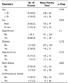
A retrospective study was conducted on 33 patients (20 men, 13 women; mean age, 67.5 years; mean interval to first stenosis, 17 months), who had performed percutaneous angioplasty for first episode of stenosis after brachioaxillary graft surgery. We evaluated the relevant factors affecting the graft patency after first episode of stenosis, such as age, sex, underlying disease (hypertension, diabetes mellitus, hyperlipidemia, cardiovascular disease, cerebrovascular attack), anastomotic angle between graft and axillary vein, and anastomotic angle between the graft and brachial artery. Kaplan-Meier method and log rank test and receiver operating characteristics curve analysis were used in statistical analysis.
Results
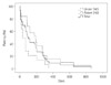
Graft patency rates after 1 month, 6 months, and 12 months were 75.8%, 39.4%, and 9.1%. There was a correlation between graft-axillary vein anastomotic angle and patency rates (r = 0.372, p = 0.033); larger the venous anastomotic angle, the longer patency rate. However, it does not come up with significant results in patency rates on age, sex, underlying disease, and graft-brachial artery angle.
Figures and Tables
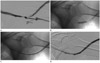 | Fig. 1Images from a 76-year-old male patient who had occlusive symptoms. It was first occlusion after arteriovenous fistula formation. He was treated for brachiaxillary type AVF graft occlusion.
A. There is stenosis and thrombosis in venous anastomosis site on angiogram. The anastomotic angle is 21°.
B. By using 6 mm-4 cm balloon catheter, balloon dilatation was performed to stenotic lesion.
C. Thrombus within the graft was completely macerated and aspirated by using Arrow-Trerotola device.
D. There is no stenosis and thrombosis in final angiogram. This patient's AVF primary patency was 19 days.
Note.-AVF = arteriovenous fistula
|
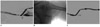 | Fig. 2Images from a 41-year-old female patient. She was treated for brachiaxillary type arteriovenous fistula graft occlusion.
A. Venous anastomotic site stenosis is shown on angiogram. The anastomotic angle is 60°.
B. Balloon dilatation was performed.
C. There is no stenosis in final venogram. Her primary patency was 650 days.
|
References
1. Bell DD, Rosental JJ. Arteriovenous graft life in chronic hemodialysis. A need for prolongation. Arch Surg. 1988. 123:1169–1117.
2. Turmel-Rodrigues L, Pengloan J, Baudin S, Testou D, Abaza M, Dahdah G, et al. Treatment of stenosis and thrombosis in haemodialysis fistulas and grafts by interventional radiology. Nephrol Dial Transplant. 2000. 15:2029–2036.
3. Butterworth PC, Doughman TM, Wheatley TJ, Nicholson ML. Arteriovenous fistula using transposed basilic vein. Br J Surg. 1998. 85:653–654.
4. Wedgwood KR, Wiggins PA, Guillou PJ. A prospective study of end-to-side vs. side-to-side arteriovenous fistulas for haemodialysis. Br J Surg. 1984. 71:640–664.
5. Kim YO, Choi YJ, Kim JI, Kim YS, Kim BS, Park CW, et al. The impact of intima-media thickness of radial artery on early failure of radiocephalic arteriovenous fistula in hemodialysis patients. J Korean Med Sci. 2006. 21:284–289.
6. Malovrh M. Non-invasive evaluation of vessels by duplex sonography prior to construction of arteriovenous fistulas for haemodialysis. Nephrol Dial Transplant. 1998. 13:125–129.
7. Kim YO, Song HC, Yoon SA, Yang CW, Kim NI, Choi YJ, et al. Preexisting intimal hyperplasia of radial artery is associated with early failure of radiocephalic arteriovenous fistula in hemodialysis patients. Am J Kidney Dis. 2003. 41:422–428.
8. Wong V, Ward R, Taylor J, Selvakumar S, How TV, Bakran A. Factors associated with early failure of arteriovenous fistulae for haemodialysis access. Eur J Vasc Endovasc Surg. 1996. 12:207–213.
9. Malovrh M. Native arteriovenous fistula: preoperative evaluation. Am J Kidney Dis. 2002. 39:1218–1225.
10. Konner K. The anastomosis of the arteriovenous fistula--common errors and their avoidance. Nephrol Dial Transplant. 2002. 17:376–379.
11. Glass C, Johansson M, DiGragio W, Illig KA. A meta-analysis of preoperative duplex ultrasound vessel diameters for successful radiocephalic fistula placement. J Vasc Ultrasound. 2009. 33:65–68.
12. Saucy F, Haesler E, Haller C, Déglise S, Teta D, Corpataux JM. Is intra-operative blood flow predictive for early failure of radiocephalic arteriovenous fistula? Nephrol Dial Transplant. 2010. 25:862–867.
13. Lin CH, Chua CH, Chiang SS, Liou JY, Hung HF, Chang CH. Correlation of intraoperative blood flow measurement with autogenous arteriovenous fistula outcome. J Vasc Surg. 2008. 48:167–172.
14. Lazarides MK, Georgiadis GS, Antoniou GA, Staramos DN. A meta-analysis of dialysis access outcome in elderly patients. J Vasc Surg. 2007. 45:420–426.
15. Rooijens PP, Tordoir JH, Stijnen T, Burgmans JP, Smet de AA, Yo TI. Radiocephalic wrist arteriovenous fistula for hemodialysis: meta-analysis indicates a high primary failure rate. Eur J Vasc Endovasc Surg. 2004. 28:583–589.
16. Culp K, Flanigan M, Taylor L, Rothstein M. Vascular access thrombosis in new hemodialysis patients. Am J Kidney Dis. 1995. 26:341–346.
17. Ku YM, Kim YO, Kim JI, Choi YJ, Yoon SA, Kim YS, et al. Ultrasonographic measurement of intima-media thickness of radial artery in pre-dialysis uraemic patients: comparison with histological examination. Nephrol Dial Transplant. 2006. 21:715–720.
18. Sedlacek M, Teodorescu V, Falk A, Vassalotti JA, Uribarri J. Hemodialysis access placement with preoperative noninvasive vascular mapping: comparison between patients with and without diabetes. Am J Kidney Dis. 2001. 38:560–564.
19. Konner K, Hulbert-Shearon TE, Roys EC, Port FK. Tailoring the initial vascular access for dialysis patients. Kidney Int. 2002. 62:329–338.
 XML Download