

 PDF
PDF ePub
ePub Citation
 Print
Print


INTRODUCTION
The prognosis of advanced gastric cancer (AGC) depends on a variety of different factors, including tumor size, depth of invasion, nodal metastases, and pathological type. Among these factors, depth of tumor invasion and nodal metastases are regarded as crucial for treatment planning, as well as determining patient prognosis (1-3). Many investigators have reported that gross classification of Borrmann type may also be a valuable clinicopathological factor. Li et al. (4) reported that the prognoses of patients with Borrmann type IV AGCs (infiltrative type) are significantly worse than those of patients with Borrmann type III AGCs (ulceroinfiltrative type), the 5-year survival rates of Borrmann types III and IV were 55.2 and 31.8%, respectively. Furthermore, presence of Borrmann type IV AGC is accepted as an independent prognostic factor of decreased patient survival (4-6). The vast majority of gastric cancers are characterized endoscopically prior to treatment; however, due to the lack of grossly defidistinguishable mucosal lesion, lesion detection and determining the extent of Borrmann type IV lesions have been challenging for endoscopists (7, 8).
Development of multi-detector row computed tomography (MDCT) has resulted in the ability to obtain improved abdominal imaging resolution with decreased scan time. In addition, the multiplanar capability of MDCT allows for very high quality stomach imaging. Currently, two-phased (arterial and portal phases) dynamic contrast-enhanced MDCT is generally used for the staging of gastric cancers (9, 10). Takao et al. (11) reported that 28% of AGCs on triphasic spiral CT demonstrate gradual enhancement, and that 73% of these tumors consist of scirrhous carcinoma. Thus, we hypothesized that there may be different enhancement patterns to characterize Borrmann IV AGCs showing the poorest prognosis from other types of AGCs, including Borrmann III lesions with literally-similar infiltrative pattern of tumor growing on different dynamic phases of contrast enhanced CT. The purpose of our study was to investigate the capability of two-phased dynamic MDCT, using the water filling method to discern between Borrmann type IV and type III AGCs by evaluating the enhancement patterns.
MATERIALS AND METHODS
Patient Populations
We obtained approval for this study from the institutional review board at our hospital, and the requirement for informed consent from individual patients was waived. After reviewing electronically stored clinical records from January 2002 to December 2005, we identified a total of 225 patients who had pathologically proven Borrmann type III (n = 160) and type IV (n = 65) AGCs. All of the pre-operative CT images were initially reviewed by a study coordinator with 10 years of experience with abdominal imaging, and 82 patients including the majority of referred patients from other hospital were excluded because they were not examined by a dual phase CT or water-filling method of gastric lumen by the standard pre-operative protocol of gastric cancer in our institution. Finally, a total of 143 patients [Borrmann type IV, n = 43 (M : F = 25 : 18, 32-75 years old, mean = 56 years old); Borrmann type III, n = 100 (M : F = 65 : 35, 21-87 years old, mean = 54 years old)] were selected for the retrospective analysis of imaging features.
CT Techniques
Two-phased (arterial and portal) contrast enhanced dynamic CT was performed with a 4-MDCT scanner (LightSpeed, GE Healthcare, Milwaukee, WI, USA) and 16-MDCT scanner (Somatom Sensation 16, Siemens Medical Solutions, Forchheim, Germany). Patients fasted for at least 6 hours before examination and then ingested between 600 to 1000 mL of water to distend the stomach immediately prior to CT examination. Because of the middle or distal gastric locations of the primary lesions, most patients were placed in prone position on the scanning table. However, patients with known primary lesions in the fundal portion of the stomach were placed in supine position. All patients received 150 mL of IV nonionic contrast material (iopromide, Ultravist 300, Bayer Health Care, Berlin, Germany) by means of a power injector (Envision CT, Medrad, Indianola, PA, USA) at a rate of 3 mL/s. Scans were acquired in a craniocaudal direction with the following parameters: detector collimation, 16 × 0.75 mm; table feed, 12 mm per rotation; section width, 5 mm; reconstruction increment, 5 mm with 5-mm-thick sections; pitch, 1.2; tube current, 120 kVp; and 160 mAs. Acquisition of arterial phase scans was initiated 15 seconds after enhancement of the thoracic aorta, until 100 Hounsfield unit was reached as measured with a bolus-tracking technique after injection of the contrast material. Portal venous phase scans were acquired 70 seconds after the start of the contrast injection.
Image Analysis
Two radiologists with 6 and 7 years of experience in abdominal CT interpretation reviewed all patient CT images without any clinical information. Before the review, Borrmann type IV lesions were presumed to exhibit layered patterns of contrast enhancement of the diseased gastric wall with time-dependant gradual thickening of an inner layer of contrast enhancement. Conversely, Borrmann type III lesions were assumed to exhibit rather inhomogeneous contrast enhancement throughout the diseased gastric wall with little difference between the arterial and portal phases. The radiologists retrospectively and independently scored tumor enhancement patterns of two-phased dynamic CT images on a picture archiving and communication system monitors using a five-grade scale. The presence or size of ulcerations was not considered as a determinant of the score for each lesion. The five-grade scale was as follows: Grade 1 - definite heterogeneous enhancement of the entire tumor lesion on arterial and portal phase images; Grade 2 - probably heterogeneous enhancement of the entire tumor lesion on arterial and portal phase images; Grade 3 - undetermined enhancement pattern of the entire tumor on arterial and portal phase images; Grade 4 - probably homogeneous enhancement of the entire tumor lesion with a gradually layered pattern of contrast enhancement on arterial and portal phase images; and Grade 5 - definite homogeneous enhancement of the entire tumor lesion with a gradually layered pattern of contrast enhancement on arterial and portal phase images.
Data Analysis
SPSS software (version 15.0; Statistical Package for the Social Science, Chicago, IL, USA) was used for statistical analyses. A weighted kappa test was applied to measure interobserver variability. The analysis of the correlation between the score of tumor enhancement pattern and the macroscopic Borrmann type was determined using Spearman's correlation coefficient. For evaluation of the accuracy of differentiation of Borrmann type IV using MDCT in each reviewer, receiver operating characteristic (ROC) analysis was used (12-14). In all statistical analyses, differences were considered significant when the p value was less than 0.05.
RESULTS
Interobserver agreement for the CT pattern scores of Borrmann types was substantial (weighted kappa = 0.683) between the two reviewers for each lesion. The grade of tumor enhancement pattern showed a significant correlation with Borrmann types (reviewer 1, r = 0.591, p < 0.001; reviewer 2, r = 0.616, p < 0.001), which suggested Borrmann type IV lesions tend to exhibit more uniformly layered enhancement patterns, while Borrmann type III lesions exhibit more inhomogeneous enhancement patterns.
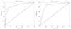
The area under the ROC curve (Az) for differentiation of Borrmann type by two-phased dynamic MDCT was 0.86 (p < 0.001) in both reviewers. Using an optimal threshold (Grades 3-5) for the diagnosis of Borrmann type IV distinguished from Borrmann type III lesions, the sensitivity and specificity were 79% and 82% in reviewer 1 and 88% and 78% in reviewer 2, respectively (Figs. 1, 2, 3, Table 1).
DISCUSSION
In Western countries, more than 80% of patients with gastric cancer have AGC, and the prognosis of gastric cancer is still unsatisfactory, even after radical gastrectomy (15). Morphological classification of AGC using the Borrmann classification system (types I through IV) was described in 1926, and since then, it has been widely used (16, 17). Borrmann type IV has shown an independent poor prognostic factor, and An et al. (5) reported that the 5-year survival rate of Borrmann type IV patients is significantly lower than that of patients with other Borrmann types (5-year survival rate on Borrmann type IV versus other Borrmann types, 27.6% versus 61.2%, respectively). At the time of diagnosis, Borrmann type IV tend to show more advanced T-stages of gastric cancer, compared to the other Borrmann types and the rate of T4 lesions are 11.9%, 6.3%, 16.4% and 30.7% in Borrmann types I, II, III and IV, respectively (6). Meanwhile, the overall incidence of Borrmann type IV is reported to represent 11-13% of all AGCs (18, 19).
Dynamic MDCT with the water filling method has several merits, such as clear depiction of the gastric wall, prominent tumor enhancement, inexpensive material, good toleration, and lack of overshooting artifacts by air in the lumen. Shimizu et al. (20) reported that the detection rate of AGC is high (96.2%) when using MDCT with the water filling method and that the accuracy of the depth of invasion of gastric cancer is also high (85%). On dynamic MDCT scan with water filling, the layered pattern of normal gastric wall and early gastric cancer are mostly visualized in the arterial dominant phase, and the depth of tumor invasion, in the case of AGC, is well verified in the equilibrium phase (11, 21). On two-phased dynamic MDCT using the water filling method, the arterial phase has a role in detection and differentiation of early gastric cancer and AGC. Likewise, the portal phase has a role in evaluation of depth of invasion (T staging). In this study, the arterial phase almost always revealed prominent enhancement of the luminal portion of AGC, while the portal phase identified depth of tumor invasion through the deeper portion of the stomach wall; thereby, allowing the determination of tumor extension by the degree and pattern of enhancement.
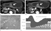
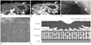
Borrmann type IV AGC tends to be pathologically defined as an infiltration into the serosal layer without any distinct elevation or crater, resulting in a rigid and thick gastric wall with giant mucosal folds. In the early stages of tumor growth in Borrmann type IV AGC, tumor cells individually invade the lamina propria without changing the mucosal surface, grow into the submucosal plane, and invade the whole stomach wall without distinct borders. Thus, Borrmann type IV lesions are not usually detected early and are generally associated with a poor prognosis (22, 23). Especially during the spread of a tumor into the submucosa and proper muscle layers of the stomach wall, tumor burdens are poorly visualized in the submucosa and muscular layers, and desmoplastic infiltrations are rather prominent (Fig. 3). Monzawa et al. (24) reported that delayed enhancement is also frequently demonstrated in tumors with abundant fibrous tissue stoma. Further, Takao et al. (11) reported that 28% of AGCs exhibit gradual enhancement on triphasic spiral CT scan, and that 73% of these tumors have pathologically demonstrated marked fibrous tissue stroma (scirrhoous carcinoma, Borrmann type IV). Based on these previous reports, we hypothesized that Borrmann type IV tumors would show early enhancement of the hypervascular superficial layer, including intact mucosal lining and subsequent enhancement of relatively hypovascular deeper layers containing abundant fibrosis with scanty tumor cells, resulting in stratification of the enhancement pattern during dynamic imaging. On the other hand, Borrmann type III lesions should have a larger cellular component with less fibrotic tumor stroma, and thus, were expected to exhibit a rather inhomogeneous contrast enhancement of the entire tumor burden depending on the degree of tumor cell infiltration across the gastric wall. In this study, Borrmann type III and IV tumors were significantly correlated with tumor enhancement pattern (diffusely heterogeneous versus homogeneous with gradually layering pattern), and were readily differentiated by enhancement pattern on a two-phased dynamic CT. Based on our results, the accuracy of correct diagnosis of Borrmann type IV by enhancement pattern on two-phased dynamic MDCT was significant (accuracy of 85%) (Fig. 1).
Our study had several limitations. First, we did not evaluate the effect of tumor enhancement by tumor histologic type or tumor grading. Borrmann type IV has a high proportion of poorly differentiated adenocarcinoma compared to Borrmann type III (6); however, Yin et al. (25) reported that an enhanced ratio on dual-phased contrast enhanced MDCT is not correlated with histo-differentiation of gastric cancer. Thus, we suggest that tumor grade has a minimal effect on tumor enhancement pattern on dynamic MDCT. Second, tumor histologic type, especially mucinous carcinoma and signet ring cell carcinoma, may have affected tumor enhancement pattern. Park et al. (26) reported that mucinous carcinoma has a layering pattern (62%) of enhancement, while Lee et al. (27) reported that signet ring cell carcinoma (SRCC) exhibits a high degree of contrast enhancement (37%). Actually we had only two cases of mucinous carcinomas (Borrmann type III, n = 1; type IV, n = 1) with corresponding enhancement patterns of our results; however, a comparison of SRCC lesions (Borrmann type III, n = 26; type IV, n = 11) showed limited specificity (sensitivity 81% and 90%, specificity 43% and 58% for reviewer 1 and 2, respectively) in the diagnosis of type IV lesions by the gradual layering pattern of mural enhancement on the dynamic CT. Considering the exceptional features of SRCC lesions, further study would be needed according to the histologic types of lesions. In addition, we did not evaluate the morphologic appearance of Borrmann types on MDCT during image analysis. Specifically, if prediction of tumor gross appearance had been evaluated, the reviewers would have had a preconception during subsequent analysis of tumor enhancement patterns. To minimize such bias, reviewers analyzed only tumor enhancement patterns on images and were not aware of any additional information. Lastly, for endoscopists, the most important clinical issue of Borrmann type IV may be lesion delineation rather than differentiation from Borrmann type III lesions; and, the impact of our present imaging study may be limited to MDCT characterization of Borrmann type IV lesions. However, our results could be helpful for the endoscopically suspicious cases of Borrmann type IV with negative endoscopic biopsy results due to little tumor burden with fibrosis at thickened gastric wall (Fig. 3).
In conclusion, two different tumor enhancement patterns of diffusely heterogeneous versus homogeneous with gradual thickening of enhancing portion (layered pattern) were considerably matched with Borrmann type III versus IV, respectively, on dual-phased dynamic MDCT using the water filling method. Based on the results of this study, with considering the exception of SRCC lesions, MDCT characteristics may offer useful information for diagnosing Borrmann type IV in AGC, which could be helpful for detection of inexpectant Borrmann type IV and suspicious Borrmann type IV with negative biopsy by endoscopy.

 XML Download