

 PDF
PDF ePub
ePub Citation
 Print
Print


INTRODUCTION
Anatomical and morphological variation of vertebral arteries is important in neurointervention, surgery and other non-invasive procedures (1). The left vertebral artery almost always arises as the first branch of the left subclavian artery. However, in approximately 5% of all cases, the left vertebral artery has been reported to arise at the aortic arch (2-4). In some of these cases, duplication of the vertebral artery occurs, although this is very rare and has only been reported a few times (2-4). Anecdotal evidence describes clinical symptoms, such as dizziness or vertigo, in patients with anomalous vertebral artery origins, although there is no conclusive evidence of an association with cerebrovascular accidents (2); there has also been much controversy over the supposed negative effects of vertebral artery duplication. An effect on the hemodynamics of the intracranial component is a possibility (2). In this study, we describe a patient with left vertebral artery duplication which was detected incidentally.
CASE REPORT
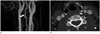
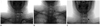
A dual origin of the left vertebral artery and an aneurysm whose origin was shared with the left anterior temporal artery were incidentally detected by magnetic resonance angiography (MRA) and computed tomography angiography (CTA) in a 51-year-old female who visited our institute for health screening (Fig. 1). After the examination, conventional angiography was performed on the patient. The anomalous vessel was discovered to be the third branch of the aortic arch, and it opacified the left vertebral artery due to the retrograde flow into the second limb, which arose as the first branch of the left subclavian artery (Fig. 2). Selective left vertebral angiography through the usual vertebral artery origin, which is the left subclavian artery, revealed retrograde flow of the left vertebral artery into a hypoplastic limb arising from the aortic arch. CTA, MRA and angiography showed that the proximal duplicated limbs of the left vertebral artery united to form the distal part of the left vertebral artery at the C4 level.
DISCUSSION
The term "vertebral artery duplication" is applied to a vertebral artery that has two origins with fusion at the neck level. The duplicated segments almost always fuse at C4-6 (5). Vertebral artery duplication occurs in cases in which an artery originates in an unusual location. A number of studies have reported the frequency of anomalous vertebral artery origins. Panicker et al. (6) reported an abnormal left vertebral artery origin in 1 out of 20 middle-aged female cadavers during the period 1998-2002. In a study by Bergman et al. (7) on 693 laboratory specimens, a left vertebral artery arising at the aortic arch between the left common carotid and left subclavian arteries was reported in 2.46% of all specimens (17 of 693). A right vertebral artery arising at the site of bifurcation of the innominate artery into the right subclavian and right common carotid arteries was reported in 1.11% of all specimens (7 of 693). However, duplication of vertebral arteries is extremely rare. Only three cases of a duplicated left vertebral artery arising from the aortic arch and the left subclavian artery have been reported (2-4). A hypoplastic duplicated limb was reported in two of these cases; in one case, it was the branch from the aortic arch and in the other, the branch from the left subclavian artery. In the case reported here, the hypoplastic limb was a direct branch from the aorta.
Right vertebral artery duplication has occasionally been reported. Goddard et al. (5) reported two cases of duplication of the right vertebral artery. In one case, the duplication was proximal, with both origins located in the right subclavian artery (the origins were not described in the other case). Satti et al. (2) also reported a case in which the right vertebral artery was partially duplicated with a common origin as the second branch of the right subclavian artery. However, a duplicated vertebral artery origin appears to be more common on the left side.
Embryologically, the vertebral arteries typically originate bilaterally from the distal end of the seventh dorsal intersegmental arteries. A duplicated vertebral artery occurs if additional cervical intersegmental branches arise from the descending aorta (2, 3). This is due to the failure of the right or left fifth intersegmental artery to regress (5).
A complete understanding of the origins of anomalous vertebral arteries may be important. Vertebral arteries almost always enter the C6 transverse foramen (8). However, in cases where the left vertebral artery arises from the aortic arch between the left common carotid artery and the left subclavian artery, it generally enters the transverse foramen at the level of C4-C5 rather than C6 (9). Therefore, the risk of atherosclerosis in vertebral arteries arising from the aortic arch may be higher than in those with other configurations due to their long prevertebral segments. In addition, a left vertebral artery of the aortic origin is thought to be associated with a higher incidence of vertebral artery dissection than a left vertebral artery of the left subclavian artery origin (10). Anomalous vertebral artery origins affect hemodynamics and may lead to intracranial malformation. In 2009, Kendi and Brace (4) reported a case of a duplicated vertebral artery with an intracranial aneurysm. However, there is no conclusive evidence that this congenital anomaly predisposes to cerebrovascular accidents (2); thus, more studies are needed to determine its consequences.
The presence of a vertebral artery segment outside the bony cervical spine as high as C4 (the level of the carotid bifurcation in the majority of patients) is important for neurointervention or spinal surgery. The knowledge of the potential left vertebral artery origin variants may influence the choice and route of the endovascular treatment (7).
In conclusion, duplication of vertebral artery origins is extremely rare, but may have serious implications, with particular regards to hemodynamics, angiography, endovascular and surgical procedures.
 XML Download