

 PDF
PDF ePub
ePub Citation
 Print
Print


INTRODUCTION
Intracranial chordomas are relatively rare, locally aggressive, malignant lesions constituting about 1% of all intracranial tumors and 4% of all primary bone tumors (1). They arise from notochordal remnants, usually at the sphenooccipital synchondrosis involving the clivus. Approximately 35-40% of these tumors occur intracranially, where they typically involve the clivus. Most clivus chordomas show extradural extension and bone destruction. Very rarely, a chordoma can arise intradurally without any bony involvement (2). This report is concerned with the radiological findings in prepontine intradural chordoma having no clival bone involvement, which is challenging in order to make a differential diagnosis.
CASE REPORT
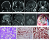
A 32-year-old man had been complaining of dysarthria for 3 years. There were no other motor, sensory, or cerebellar deficits. CT revealed a hypodense pontine mass without contrast enhancement and there was no bony erosion at the clivus (Fig. 1A). MRI showed an extrinsic multilobulated contour-bulging lesion in the prepontine cistern mildly extending into the right cerebellopontine angle and invaginating into the pontine parenchyma. This mass partially encased the mid basilar artery. The mass was hypointense on T1-weighted and hyperintense on T2-weighted images. Most of the mass showed no post-contrast enhancement except some small central multiple reticulonodular heterogenous enhancements. There was no demonstrable bony erosion or destruction of the clivus with preserved normal fatty marrow (Fig. 1B-F). On the diffusion-weighted images (DWI), this mass showed heterogenous subtle increased signal intensity (Fig. 1G). Preoperative diagnosis was considered as an epidermoid cyst, most likely due to low T1-weighted and high T2-weighted signal intensity. On post-contrast enhanced magnetic resonance (MR) images, the presence of multifocal intratumoral small enhancing foci, which was not an usual findings of epidermoid cyst, was suggested as the intratumoral fine multiple atypical vascular foci or early malignant transformation in the epidermoid cyst. However, on DWI images, the degree of high signal intensity was not a classic feature of an epidermoid cyst.
The patient underwent an extended trans-sphenoidal approach and subtotal tumor removal. At surgery, the mass was in purely intradural location, with pale grayish gelatinous and glistening feature (Fig. 1H). At surgical field, there was a suspicious small defect area in the retroclival prepontine dura. The diagnosis of chordoma was confirmed by an immunohistochemical study, as tumor cells were positive for S-100 and pancytokeratin (Fig. 1I-K). The central enhanced foci in the intradural chordoma were confirmed not as malignant transformation but as the prominent vascularity. No symptoms existed including dysarthria after the first operation. After an 8-month follow- up, since the remnant tumor was slightly growing, a second operation was undergone.
DISCUSSION
Chordoma, an uncommon neoplasm believed to originate from remnants of the primitive notochord, is found predominantly in the sacrococcygeal and spheno-occipital areas. Intracranial chordomas, which constitute approximately 35-40% of chordomas, are extradural tumors arising mostly at the sphenooccipital synchondrosis in the clivus. Rarely, they arise in the sella, sphenoid sinus, nasopharynx, maxilla, and paranasal sinuses (23). These tumors are slowly growing and locally invasive, and cause extensive bone destruction. After intravenous injection of contrast medium, chordomas usually enhance homogeneously or heterogeneously (4). MRI shows a low signal lesion on T1-weighted images, and a heterogeneous or homogeneous high signal on T2-weighted images with heterogenous mild to moderate contrast enhancement. Also, areas of hemorrhage, calcification, and proteinacious accumulation can be seen (5). Intradural chordomas are very rare. The theory regarding intradural chordomas is that some notochordal remnants arising from the clivus are supposed to be departed from the main part of the notochordal remnant and moved into the dura and entrapped in the intradural space. The invagination of the pontine parenchyma in our case is probably due to the slow growth within the limited intradural space.
Nishigaya et al. (6) in a review of literature regarding intradural chordomas identified only 14 such cases in 1998. Following this, around four other cases have been reported (7). Our case represents another example of an intradural chordoma, unique in that full radiological studies, including CT, MRI and cerebral angiography, were conducted. Also, the tumor was attached to the clivus without destroying it.
The histological differential diagnoses for this lesion are ecchordosis physaliphora, chordoid meningiomas, or metastatic mucinous carcinomas (36). In addition, our case should be made a differential diagnosis in addition, our case should be made a differential diagnosis from the epidermoid cyst. from the epidermoid cyst. The epidermoid cyst is a congenital cystic lesion that arises from epithelial inclusions at the time of neural tube closure or during formation of the secondary cerebral vesicles (8). It is a benign extracerebral intradural lesion and in about 40% of cases is located in the cerebellopontine angle. Due to their avascular nature and composition with cholesterol in a solid crystalline state and keratin within the tumor, epidermoid tumors typically have low signal intensity on T1-weighted images, high signal intensity on T2-weighted images, and no enhancement on gadolinium-enhanced images. However, epidermoid tumors showing unusual signal intensity changes have been reported caused by high protein concentration or an intracystic hemorrhage. These rare cases have high signal intensity on both T1-weighted and T2-weighted images and a high T1-weighted and low T2-weighted images due to the mixture of keratin with differently aged hemorrhage and granulation tissue changes. Furthermore, they showed an obvious enhanced part on contrast- enhanced T1-weighted images (8). However, these tumors show hyperattenuation on CT scan, and bright signal intensity on DWI, which is not consistent with our case. Ecchordosis physaliphora is an ectopic notochord tissue found intradurally, anterior to the pons in the prepontine cistern. It is attached to the dorsal clivus via a cartilaginous or osseous stalk (3). Moreover, the histological and immunohistochemical features of ecchordosis physaliphora and intradural chordoma are identical. Both are positive for S-100 and cytokeratin. Electron microscopic studies have also shown no significant ultrastructural differences (6). However, the differentiating points are that, unlike chordomas, ecchordosis physaliphora is usually asymptomatic and does not enhance on contrast MRI images (39). Intradural chordomas also have to be differentiated from chordoid meningiomas. Chordoid meningioma is a rare subtype of meningioma, which shares similar MRI features as meningioma. Diagnosis is established by cytopathology. Meningiomas are S-100 negative, and vimentin positive (10). Adenocarcinomas can be differentiated by the absence of S-100 and vimentin staining.
With the available data at present, completely excised intradural chordomas require only follow-up with no adjuvant therapy. Nishigaya et al. (6) reported that the residual tumor showed no sign of regrowth despite the fact that radiation therapy had not been used five years after subtotal removal. Our patient didn't have adjuvant therapy after the subtotal removal. In view of the slightly increased size of the remnant intradural chordoma during 8 months following the first operation, the remnant tumor should be in need of additional therapy. Our patient underwent secondary surgery, and had radiation therapy. During four months after the second operation, the remnant tumor size did not change. Thus, adjuvant therapy after subtotal resection of the intradural chordoma may be indispensable. Conventional radiation therapy, stereotactic radiosurgery and proton beam therapy are alternatives (6).
We have reported a rare case of an intradural prepontine chordoma. A differential diagnosis regarding intradural chordomas must be considered when the preoperative MRI features are not consistent with an epidermoid cyst if there are multiple fine enhancing lesions on enhanced MR images and no bright signal intensity on DWI.
 XML Download