

 PDF
PDF ePub
ePub Citation
 Print
Print


INTRODUCTION
Imaging studies play a major role in the diagnosis of radial head or neck fractures. Radius fracture type is classified according to imaging findings, including the degree of fracture, displacement, the number and size of fracture fragments, and the extent of articular involvement, using Modified Mason criteria (1962) (1). Assessment of fracture type is very important for the exact diagnosis and appropriate treatment, because it determines further treatment in radial head or neck fracture (23456).
For radiographic evaluation of radial head or neck fracture, many previous efforts have been made to increase the diagnostic accuracy by performing additional radiographic views, such as the radio-capitellar view or the internal oblique or external oblique views, alongside standard radiographic views, such as the anterior posterior and lateral views (78). Computed tomography (CT) is generally thought to have higher diagnostic accuracy than radiography for detecting fracture. Multiplanar reformation images by multidetector computed tomography (MDCT) can offer more accurate information concerning the fracture, especially at complicated joints, so MDCT is commonly used as an additional imaging modality for diagnosing radial head or neck fracture. However, there have been few previous studies addressing the diagnostic accuracy of MDCT in radial head or neck fractures (910). Therefore, the purpose of this study is to evaluate the diagnostic value of MDCT in radial head or neck fracture comparing with the radiography, and to evaluate the factors that affect the image quality of MDCT.
MATERIALS AND METHODS
Materials
Institutional Review Board approval was obtained for this retrospective study and informed consent was waived. From January 2008 to June 2011, a total of 174 patients with post traumatic elbow symptoms, such as pain, swelling or bruising underwent both radiography and MDCT for evaluation. Among these 174 patients, 35 patients in whom there was no evidence of fracture were excluded. Then, 27 patients with proximal ulnar fractures and 39 patients with isolated distal humeral fractures were excluded. Among the remaining 73 patients, who were diagnosed with radial head or neck fracture, 8 patients were excluded, who were not treated or followed up at our center. Therefore, final 65 patients with radial head or neck fractures were included. One patient had bilateral radial head fracture, so finally, 66 cases of radial fractures (29 right elbows, 37 left elbows) were included for this study. The mean age was about 47 years old (range, 18-82 years) and there were 31 men and 34 women. There were 20 cases of isolated radial head, 12 cases of isolated radial neck fractures, and 34 cases of both radial head and neck fractures. Coexistent ulna fractures are noted for 15 cases and humeral fractures are noted at 5 cases. Both ulna and humeral fractures were noted with radial head or neck fracture at 3 cases.
The final diagnosis of radial fracture was made by an orthopedic surgeon, according to the patient's clinical symptom, initial and follow-up imaging study results and operative findings.
Image Techniques
In the radiographic evaluation of radial head or neck fracture, anterior posterior, lateral, internal oblique and external oblique views were performed. MDCT was performed with a 16 or 64 474channel CT scan (Somatom Sensation 16; Siemens Medical Solutions, Forchheim, Germany and Sensation 64, Siemens, Erlangen, Germany). Axial, coronal and sagittal multiplanar reformation images were made with a 2 mm slice thickness. Three dimensional images were made using the volume rendering technique (VRT) with 1 mm slice thickness and 0 mm reconstruction interval. The mean Kernal value was about B69.2f (B60-70f), the mean tube voltage was about 120.9 kVp (120-140 kVp), and the mean tube current was about 79.3 mAs (60-200 mAs). The mean time interval between radiography and MDCT was about 0.6 days (range, 0-10 days). During MDCT scanning, patient arm positioning and the flexion angle of the elbow joint was determined with at least patient's discomfort.
Image Analysis
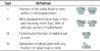
Two radiologists who were blinded to the final diagnosis and results of other imaging findings, retrospectively, reviewed the initial radiography and MDCT scans. We analyzed the presence of fracture in 66 elbows on radiography and MDCT, including VRT images and the detection rate of fracture, were calculated in both imaging tools. Further, we analyzed the fracture type in 60 elbows, excluding 6 post reductional MDCT, and compared these results with the final diagnosis. The fracture type was divided into four groups, according to the Modified Mason criteria (1) (Table 1). All image analysis was done by using images displayed on computer monitors with Picture Archiving and Communication System.
In order to evaluate the MDCT image quality, we divided patients into two groups: the good image quality group (Group A) and the poor image quality group (Group B), through a consensus procedure of the two radiologists. For this analysis, we scored each MDCT scan on a 4-point scale, 1 being poor and nondiagnostic; 2 being fair, could be nondiagnostic or confused; 3, good, diagnostic; and 4, excellent. An excellent (score of 4) was assigned when the image was 100% sharp and virtually free of degradation and background noise. Fracture sites were clearly visible in this group without any ambiguity. Fair (a score of 2) represented a blurring of the radius cortex and trabeculation of cancellous bone, which may result in nondiagnostic or confused images. A poor image quality (score of 1) did not allow evaluation of the fracture lines or fragments that are necessary for diagnosis. Good (score of 3) represented a partially blurred radius cortex and trabeculation of cancellous bone, and was assigned on the basis of the radiologists' subjective judgment between fair (score of 2) and excellent (score of 4) image quality, provided the image was still diagnostic. Patients that scored 3 or 4 were included in Group A (good image quality), and those who scored 1 or 2 were included in Group B (poor image quality).
The factors that were thought to affect the image quality of MDCT were patient's arm positioning, the flexion angle of the elbow joint, immobilization, and cancellous bone density. Arm positioning of the patient was determined by patient himself during the examination, which makes patient more comfortable with lesser pain. The flexion angle of the elbow joint was measured by drawing an extension line of the radial axis and humeral axis on the sagittal plane of MDCT. Whether immobilization is needed or not before the MDCT scanning was determined by the emergency department or orthopedic doctors who examined patients at the emergency room. Cancellous bone density was measured at the proximal radius avoiding the fracture site, using Hounsfield units (HU), which was measured by using a region of interest of 25 mm2 on sagittal plane.
Statistical Analysis
Kappa statistics was used for analyzing an inter-observer reliability between the two radiologists. A kappa value was categorized as slight (0-0.20), fair (0.21-0.40), moderate (0.41-0.60), substantial (0.61-0.80), and almost perfect (0.81-1.00). The detection rate of fracture between radiography and MDCT (Pearson's chi-square test) and the concordant rate of fracture types and the analysis of the concordant rate between the final diagnosis and each imaging modality (Kendall's tau-b value, Gamma value) was conducted using a cross tabulation. When analyzing the factors that affect MDCT image quality, the flexion angle of the elbow joint and cancellous bone density were analyzed by using independent samples t-test and patient arm positioning, and immobilization were analyzed by cross tabulation. p values under 0.05 were thought to be statistically significant and SPSS software (ver. 17.0 for Windows; SPSS Inc., Chicago, IL, USA) was used for analysis.
RESULTS
Inter-observer reliability between two readers in detecting fracture was almost perfect on both radiography (kappa value = 0.841), and MDCT (kappa value = 1.000). In addition, inter-observer reliability between two readers in classification of fracture types was almost perfect on both radiography (kappa value = 0.898) and MDCT (kappa value = 0.935). For narrowing the different opinion of two readers in some cases, readers discussed together and then made final agreement for analyzing.
In our study, radial fractures were noted in 63 of the 66 cases (95.5%) using MDCT, and in 58 cases (87.8%) using radiography. There were 8 cases (12.2%) which were detected only on MDCT and 3 cases (4.5%) which were detected on radiography, which were missing on MDCT. However, there was no statistically significant difference in the detection rates of fracture between the two imaging modalities (p = 0.065). Among the 8 missing cases by radiography, 5 cases had a cortical fracture without displacement, 2 cases had a fracture line obscured by proximal ulna, and 1 case had a fracture line obscured by osteophytes of radius. Among the 3 missing cases by MDCT, 2 cases had cortical fractures without displacement, and 1 case had a transverse radial neck fracture.
On radiography, 50 cases (75.8%) of radial head or neck fractures were detected by anterior posterior and lateral views, however, 58 cases (87.8%) were detected by an additional internal and external oblique views.
On analyzing the fracture types, 38 cases were matched between radiography and MDCT, but 22 cases were not matched between the two imaging modalities (kappa value = 0.489) (Table 2). The fracture type was accurately diagnosed in 56 cases among 60 cases with MDCT (93.3%) (Kendall's tau-b value = 0.960, Gamma value = 1.000), and in 48 cases with radiography (70.0%) (Kendall's tau-b value = 0.799, Gamma value = 0.960). The diagnostic accuracy of fracture type was significantly higher with MDCT as compared to that of radiography (p < 0.0001).
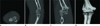
Among the 22 mismatched cases of radial fractures, except 11 cases, which were only detected either radiography or MDCT, 5 cases were thought to be type I on radiography but type II on MDCT. Remaining 6 cases were thought to be type II fracture on radiography but diagnosed as type III fracture on MDCT (Fig. 1).
There were 8 cases that were initially diagnosed as type IV fracture on radiography. Two cases were also noted as type IV on MDCT and other 6 cases were not evaluated by MDCT because closed reduction of elbow joint was performed before MDCT scanning, as mentioned before.
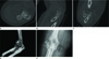
Twenty-seven cases were included in group A, and 39 cases were included in group B. All of the 8 cases, which were only detected by MDCT, were included in group A, and all of the 3 cases that were not detected by MDCT, but were detected by radiography, were included in group B. Considering the factors that were thought to affect the image quality of MDCT, there were 22 cases in which patients had their arms raised in group A, and 4 cases in group B. There were 35 cases with a lower arm position in group B and 5 cases in group A. The difference in arm positioning showed a significant difference between group A and B (p < 0.0001). The mean flexion of the elbow joint was significantly lesser in group A (about 51.9 ± 27.0°) than it was in group B (about 69.6 ± 20.8°) (p = 0.004). The mean cancellous bone density was significantly higher in group A (345 ± 74.5 HU) than it was in group B (288.8 ± 91.0 HU) (p = 0.010) (Figs. 2, 3). Immobilization was done in 10 cases in group A and 23 cases in group B, and there was no significant difference between the two groups (p = 0.132) (Table 3).
DISCUSSION
Radial head or neck fracture is the most common fracture among injuries of the elbow joint, which accounts for 1.7 to 5.4% of all fractures (6) and 25 to 44% of all elbow joint fractures (4). In our study, the detection rate for radial fracture was slightly higher using MDCT (95.5%) than radiography (87.8%), but there was no significant difference between these two modalities (p = 0.065). In a previous study by Chapman et al. (11), the detection rate of MDCT for radial fractures of children was 95.5%, which resembled similar to our study. However, the detection rate for radial fracture on MDCT is slightly lower than the detection rate for other site fractures, such as the pelvic bone, humerus, carpal bones and scapula, which showed almost about 100% accuracy with MDCT (91012131415). So, radiography can remain as the primary imaging modality to evaluate radial head or neck fracture for patients with elbow trauma.
Dillon et al. (8) noted that the external oblique view is helpful for increasing the reproducibility of a fracture type diagnosis. In this study, we did not specifically evaluate the relationship between the fracture type and different radiographic views, but the detection rate of fracture increased by using additional internal and external oblique views with routine anterior posterior and lateral views. However, the 8 cases remained negative radiographic findings, though additional internal, external oblique views were done.
However, during the study, there noted 8 missing cases that were not seen on radiography, but seen on MDCT. Among those missing cases, there were 5 cases with small cortical fracture without displacement and 2 cases with overlapping other components of the elbow joint, which made it difficult to detect fracture lines. In one case, fracture lines were masked by osteophytes associated with degenerative osteoarthritis.
Among the many methods for defining radius fracture types, the Modified Mason criteria was thought to be the most reliable method based on previous studies (124). Treatment of radial fracture is determined by the fracture type: type I and II fractures are treated with a cast or splint immobilization, whereas type III and IV fractures are treated with operative treatment, such as an open reduction with internal fixation or radial head orthoplasty. Therefore, accurate classification between type II and type III fractures is very important in clinical practice (3). MDCT with multiplanar images is a useful method for detecting small size fracture fragments and diagnosing type III fracture. In this study, the accurate diagnosis of fracture type was significantly higher using MDCT, especially in the 6 cases which were misdiagnosed as type II fracture by radiography, and were finally diagnosed as type III fracture by MDCT, after then they underwent operative treatment. Further, in another 5 cases, there noted combined displacement with fracture on MDCT, which was not definite on radiography. In those cases, there made different fracture type between type I on radiography and type II on MDCT. Otherwise, in those cases, treatment plan was not changed. So for a more accurate diagnosis of the fracture type, additional MDCT can be helpful.
However, there were 3 missing cases that were not diagnosed on MDCT. Among them, 2 cases had small cortical fractures without displacement of fracture fragment. In those cases, image quality of MDCT was too poor to detect fracture. In addition, another 1 case was radial neck fracture with fracture line parallel to the axial plane of MDCT. Further, this fracture was not seen at other plane of the MDCT.
VRT images are thought to be preferred when diagnosing small fracture displacement and for detecting fracture fragments, especially at complicated anatomical sites (16). In this study, VRT images were diagnostic for 39 cases (62.9%). The quality of VRT images is improved when the scan thickness is reduced and the reconstruction interval is shorter (16). When reconstructing VRT images, pseudolesions that have similar density to the radial head or neck can be artificially included or excluded in the final VRT images. If these are included, severe distortion can make a difficulty in the diagnosis of fracture type, but if they are excluded, useful information about the fracture can be lost.
Multiple factors are known to affect image quality in MDCT, including spatial resolution, contrast resolution, temporal resolution, CT number accuracy, noise, radiation dose and artifacts. Adjustment of these factors can improve image quality of MDCT. However, these factors are intricately connected, and factors such as scan time and scan rate are often readily fixed by the CT scan machine itself; as such, it can be hard to modify these settings for each scanning (1718192021). Tube voltage and current can also affect the image quality in MDCT, but decreasing the tube voltage can enhance the beam hardening artifact and decreasing the tube current can enhance noise, which finally yields a poor image quality (2122).
In this study, we focused on the modifiable and flexible factors that can affect image quality in MDCT, including patient arm positioning, the flexion angle of the elbow joint and whether or not immobilization was conducted. More patients raised their arms in group A; whereas, more patients lowered their arms in group B. If a patient lowers his arm during examination, his arm becomes positioned by the trunk and so the amount of X-ray that reaches the target area of the patient's elbow joint decreases (photon starvation artifact), and artifacts that are made by the patient's trunk can ultimately reduce the image quality (beam hardening artifacts) (2324). Raising the patient's arm over his head during examination is required for good MDCT image quality. If there are challenges with raising the arm at prone positioning, supine positioning may be helpful for reducing patient discomfort. In this study, image quality was notably better for the group with a lesser flexion angle of the elbow joint. When the flexion angle is increased, the humerus and radius are not located at the longitudinal parallel position; therefore, there can be artifacts caused by the humerus (beam hardening artifacts). Also, determining the axis of the coronal and axial planes is difficult. There was no significant difference found between the two groups by immobilization. The immobilization angle of the elbow joint is generally near 90°, because near this angle, there is better stabilization of the elbow joint and discomfort in patient's daily activity is decreased (25). However, if immobilization is carried out near this angle, this can result poor MDCT image quality. Consequently, considering both the effect of immobilization and good image quality, MDCT scanning is recommended to take place before immobilization of the elbow joint. When cancellous bone density is decreased, it can be hard to detect fracture lines on MDCT due to sparse trabeculation. However, cancellous bone density is a fixed patient factor that cannot be immediately modified. As such, when examining a patient with decreased cancellous bone density, follow- up or re-examination must carefully take not to misdiagnose. In summary, for better image quality and more accurate diagnosis, MDCT scanning with a raised arm position and the maximum extension of the elbow joint is recommended. Given that immobilization of the elbow joint can affect the flexion angle of the elbow joint during examination, and immobilization is recommended to take place after MDCT examination.
The limitations of this study are as follows. First, this study was designed as a retrospective study. This means that we could not confirm the results after assessing the factors that may affect MDCT image quality. Likewise, we could not compare the results for the same patients by controlling other factors. Second, the total patient number was small and patients without radial fracture were not included, so evaluation of general diagnostic accuracy was impossible.
In conclusion, the detection rate for radial head or neck fracture was slightly higher, using MDCT, compared to radiography, but there were no significant differences between these two modalities. Thus, the radiography still remains as first screening tool of the radial head or neck fracture. However, diagnostic accuracy of fracture type was noted significantly higher on MDCT than radiography. The arm positioning, flexion angle, and cancellous bone density affect MDCT image quality. For a better image quality of MDCT, scanning with a raised arm position and the smaller flexion angle of the elbow joint is recommended.


 XML Download