

 PDF
PDF ePub
ePub Citation
 Print
Print


Abstract
Apocrine carcinoma in situ of breast is a rare, unique, and morphologically distinct type of breast carcinoma. Low-grade apocrine ductal carcinoma in situ (DCIS) and apocrine metaplasia with atypia are the pathologic spectrum of apocrine breast lesions. Differentiating these two lesions is difficult due to partial microscopic overlap. We describe a case of apocrine DCIS which presented an asymptomatic hypoechoic mass with morphological change on a follow up ultrasonography.
Figures and Tables
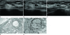 | Fig. 1A 60-year-old asymptomatic woman.
A. Initial breast ultrasonography revealed a 0.7 × 0.3 × 0.3 cm sized well-circumscribed hypoechoic mass in the left breast in the direction of 3-o'clock approximately 1 cm from the nipple.
B, C. Follow up ultrasonography after 1.5 year shows morphologic change with a newly developed indistinct margin, irregular shape and mild echogenic halo (B: longitudinal image, C: transverse image).
D. Photomicrograph shows apocrine ductal carcinoma in situ characterized by abundant eosinophilic cytoplasm and moderate nuclear pleomorphism (Hematoxylin and Eosin, × 100).
E. Photomicrograph of specimen shows a myoepithelial layer around the well-defined island of malignant cells. The neoplastic cells are negative (high molecular weight cytokeratin, × 200).
|
References
1. O'Malley FP, Bane A. An update on apocrine lesions of the breast. Histopathology. 2008. 52:3–10.
2. Visscher DW. Apocrine ductal carcinoma in situ involving a sclerosing lesion with adenosis: report of a case. Arch Pathol Lab Med. 2009. 133:1817–1821.
3. ACR BI-RADS-ultrasound. ACR breast imaging reporting and data system, breast imaging atlas. 2003. Reston, Va: American College of Radiology.
4. Doshi DJ, March DE, Crisi GM, Coughlin BF. Complex cystic breast masses: diagnostic approach and imaging-pathologic correlation. Radiographics. 2007. 27:Suppl 1. S53–S64.
5. Ogiya A, Horii R, Osako T, Ito Y, Iwase T, Eishi Y, et al. Apocrine metaplasia of breast cancer: clinicopathological features and predicting response. Breast Cancer. 2010. 17:290–297.
6. Seidman JD, Ashton M, Lefkowitz M. Atypical apocrine adenosis of the breast: a clinicopathologic study of 37 patients with 8.7-year follow-up. Cancer. 1996. 77:2529–2537.
7. Wells CA, McGregor IL, Makunura CN, Yeomans P, Davies JD. Apocrine adenosis: a precursor of aggressive breast cancer? J Clin Pathol. 1995. 48:737–742.
8. Warner JK, Kumar D, Berg WA. Apocrine metaplasia: mammographic and sonographic appearances. AJR Am J Roentgenol. 1998. 170:1375–1379.
9. Kim DY, Kang SS, Ji EK, Kwon TH, Park HL, Shim JY. Sonographic and mammographic features of breast apocrine metaplasia. J Korean Soc Ultrasound Med. 2008. 27:35–40.
10. O'Malley FP, Bane AL. The spectrum of apocrine lesions of the breast. Adv Anat Pathol. 2004. 11:1–9.
 XML Download