

 PDF
PDF ePub
ePub Citation
 Print
Print


Abstract
Purpose
To compare the linear blending image with the nonlinear moidal blending image using dual energy CT for the evaluation of the viable portion of hepatocellular carcinoma (HCC) after transcatheter arterial chemoembolization (TACE).
Materials and Methods
One-hundred and twenty three HCC patients incompletely treated after TACE were enrolled in this study. The dual energy mode (80 kVp and Sn140 kVp) was only applied in the late arterial phase scanning. A paired t-test was used to compare the lesion-to-liver contrast-to-noise ratio (CNR) and the image noise between the two blending images. Lesion conspicuity, image sharpness, image noise and the overall image quality between the two blending images were compared using the Wilcoxon matched-pair signed-ranks test.
Results
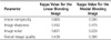
The lesion-to-liver CNR was significantly higher on the moidal blending image (5.6 ± 3.2) than on the linear blending image (2.7 ± 1.6) (p < 0.001). The image noise was significantly lower on the moidal blending image (10.9 ± 3.5) than on the linear blending image (17.5 ± 5.5) (p < 0.001). The lesion conspicuity and overall image quality were significantly better on the moidal blending image for both reviewers (p < 0.001). However, with respect to image sharpness, the linear blending image was significantly better for both reviewers (p > 0.01).
Figures and Tables
Fig. 1
A 59-year-old man who underwent two sessions of transcatheter arterial chemoembolization for HCC.
A. The unenhanced CT image shows a sparsely iodized oil-retaining lesion at the right hepatic dome (arrows).
B. The linear blending image obtained during the late arterial phase shows a questionable area of enhancement (arrows) within a sparsely iodized oil-retaining lesion at the right hepatic dome.
C. The moidal blending image obtained during the late arterial phase more clearly demonstrates the hypervascular enhancing portion (arrows) within a sparsely iodized oil-retaining lesion at the right hepatic dome.
D. The celiac axis angiogram reveals hypervascular staining (arrows) fed by branches of the right hepatic artery at the right hepatic dome.
Note.-HCC = hepatocellular carcinoma
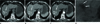
Table 1
Comparison of the Attenuation Values of the Lesions and Liver Parenchyma, the Image Noise and the Lesion-to-Liver CNR between the Linear Blending and Moidal Blending Images


References
1. Kim KW, Lee JM, Choi BI. Assessment of the treatment response of HCC. Abdom Imaging. 2011. 36:300–314.
2. Kim S, Mannelli L, Hajdu CH, Babb JS, Clark TW, Hecht EM, et al. Hepatocellular carcinoma: assessment of response to transarterial chemoembolization with image subtraction. J Magn Reson Imaging. 2010. 31:348–355.
3. Lim HS, Jeong YY, Kang HK, Kim JK, Park JG. Imaging features of hepatocellular carcinoma after transcatheter arterial chemoembolization and radiofrequency ablation. AJR Am J Roentgenol. 2006. 187:W341–W349.
4. Yu JS, Kim JH, Chung JJ, Kim KW. Added value of diffusion-weighted imaging in the MRI assessment of perilesional tumor recurrence after chemoembolization of hepatocellular carcinomas. J Magn Reson Imaging. 2009. 30:153–160.
5. Llovet JM, Real MI, Montaña X, Planas R, Coll S, Aponte J, et al. Arterial embolisation or chemoembolisation versus symptomatic treatment in patients with unresectable hepatocellular carcinoma: a randomised controlled trial. Lancet. 2002. 359:1734–1739.
6. Lo CM, Ngan H, Tso WK, Liu CL, Lam CM, Poon RT, et al. Randomized controlled trial of transarterial lipiodol chemoembolization for unresectable hepatocellular carcinoma. Hepatology. 2002. 35:1164–1171.
7. Belghiti J, Carr BI, Greig PD, Lencioni R, Poon RT. Treatment before liver transplantation for HCC. Ann Surg Oncol. 2008. 15:993–1000.
8. Kubota K, Hisa N, Nishikawa T, Fujiwara Y, Murata Y, Itoh S, et al. Evaluation of hepatocellular carcinoma after treatment with transcatheter arterial chemoembolization: comparison of Lipiodol-CT, power Doppler sonography, and dynamic MRI. Abdom Imaging. 2001. 26:184–190.
9. Jang KM, Choi D, Lim HK, Lim JH, Lee JY, Lee WJ, et al. Depiction of viable tumor in hepatocellular carcinoma treated with transarterial chemoembolization: multiphasic helical CT with review of the previous serial CT images. Korean J Radiol. 2005. 6:153–160.
10. Lau WY, Lai EC. Hepatocellular carcinoma: current management and recent advances. Hepatobiliary Pancreat Dis Int. 2008. 7:237–257.
11. Behrendt FF, Schmidt B, Plumhans C, Keil S, Woodruff SG, Ackermann D, et al. Image fusion in dual energy computed tomography: effect on contrast enhancement, signal-to-noise ratio and image quality in computed tomography angiography. Invest Radiol. 2009. 44:1–6.
12. Fletcher JG, Takahashi N, Hartman R, Guimaraes L, Huprich JE, Hough DM, et al. Dual-energy and dual-source CT: is there a role in the abdomen and pelvis? Radiol Clin North Am. 2009. 47:41–57.
13. Kim KS, Lee JM, Kim SH, Kim KW, Kim SJ, Cho SH, et al. Image fusion in dual energy computed tomography for detection of hypervascular liver hepatocellular carcinoma: phantom and preliminary studies. Invest Radiol. 2010. 45:149–157.
14. Macari M, Spieler B, Kim D, Graser A, Megibow AJ, Babb J, et al. Dual-source dual-energy MDCT of pancreatic adenocarcinoma: initial observations with data generated at 80 kVp and at simulated weighted-average 120 kVp. AJR Am J Roentgenol. 2010. 194:W27–W32.
15. Marin D, Nelson RC, Samei E, Paulson EK, Ho LM, Boll DT, et al. Hypervascular liver tumors: low tube voltage, high tube current multidetector CT during late hepatic arterial phase for detection--initial clinical experience. Radiology. 2009. 251:771–779.
16. Robinson E, Babb J, Chandarana H, Macari M. Dual source dual energy MDCT: comparison of 80 kVp and weighted average 120 kVp data for conspicuity of hypo-vascular liver metastases. Invest Radiol. 2010. 45:413–418.
17. Sommer CM, Schwarzwaelder CB, Stiller W, Schindera ST, Heye T, Stampfl U, et al. Dual-energy computed-tomography cholangiography in potential donors for living-related liver transplantation: initial experience. Invest Radiol. 2010. 45:406–412.
18. Coursey CA, Nelson RC, Boll DT, Paulson EK, Ho LM, Neville AM, et al. Dual-energy multidetector CT: how does it work, what can it tell us, and when can we use it in abdominopelvic imaging? Radiographics. 2010. 30:1037–1055.
19. Bruix J, Sherman M. American Association for the Study of Liver Diseases. Management of hepatocellular carcinoma: an update. Hepatology. 2011. 53:1020–1022.
20. Holmes DR 3rd, Fletcher JG, Apel A, Huprich JE, Siddiki H, Hough DM, et al. Evaluation of non-linear blending in dual-energy computed tomography. Eur J Radiol. 2008. 68:409–413.
21. Schindera ST, Nelson RC, Mukundan S Jr, Paulson EK, Jaffe TA, Miller CM, et al. Hypervascular liver tumors: low tube voltage, high tube current multi-detector row CT for enhanced detection--phantom study. Radiology. 2008. 246:125–132.
22. Graser A, Johnson TR, Chandarana H, Macari M. Dual energy CT: preliminary observations and potential clinical applications in the abdomen. Eur Radiol. 2009. 19:13–23.
23. Paul J, Bauer RW, Maentele W, Vogl TJ. Image fusion in dual energy computed tomography for detection of various anatomic structures--effect on contrast enhancement, contrast-to-noise ratio, signal-to-noise ratio and image quality. Eur J Radiol. 2011. 80:612–619.
 XML Download