

 PDF
PDF ePub
ePub Citation
 Print
Print


INTRODUCTION
Omphalocele is a rare congenital anomaly, in which the infant's intestines protrude through the navel. The incidence is approximately 1 : 3000-1 : 10000 live births. Additional anomalies associated with omphalocele are presented in as many as 50% of cases and vary greatly among patients (1). The most common anomalies include chromosomal defects, cardiac, genitourinary, craniofacial and diaphragmatic anomalies, which have been reported elsewhere (123). However, the persistence of these anomalies and the positioning of visceral solid organs in post-operative omphalocele patients have not been reported previously.
Herein, we report a case of post-operative omphalocele patient with abnormal positioning of the liver, spleen and both kidneys, along with anomalous drainage of the hepatic vein, which was found during a routine computed tomography (CT) scan of the abdomen and pelvis.
CASE REPORT
A parturient woman, who had not undergone gynecological examination during her pregnancy, visited the emergency room for the delivery of her child. Antenatal fetal ultrasonography showed abnormal bowel content and herniation. An emergency cesarean section was performed, and the infant was found to have a large omphalocele. By postnatal gestational age assessment, a baby was confirmed to be 42 weeks of gestational age, and her birth weight was 3150 g. The size of omphalocele was about 15 cm, and partial rupture of membrane was accompanied.
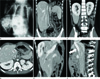
Initial infantogram (Fig. 1A) showed multiple bowel loop herniation into a membrane-covered sac. The radiopaque portion within the sac was thought to represent herniated liver and spleen. On the following day, temporary silastic silo was placed and the small and large bowel, stomach, spleen and liver were found to protrude through the abdominal wall defect. The size of abdominal wall defect was about 7 cm, and malrotation of the small and large bowels was associated. The intestines were placed in a temporary silastic silo for 1-month, and the postoperative course was uneventful. During her hospitalization, echocardiography and chromosomal study were performed to evaluate the associated anomalies. The chromosomal study was normal. In echocardiography, 4 mm sized small atrial septal defect was found. However, follow up echocardiography demonstrated a spontaneous closure of atrial septal defect at 4 month of her age.
Six years later, a follow-up abdomen and pelvis CT was performed to search for an incisional or ventral hernia. Although there was no evidence of a hernia, unexpected abnormal findings were obtained. The liver was found in a craniocaudal position, and about half of the inferior part of the liver was just beneath the abdominal wall, and not protected by the rib cage (Fig. 1B). Both kidneys were located in the upper portion of the liver and spleen (Fig. 1C). Liver, spleen and both kidneys were shown on the same level, just beneath the diaphragm (Fig. 1D). The longitudinal dimension of the liver and spleen was 9.0 cm and 6.5 cm, respectively, within normal range for her age. In case of hepatic vein, three major hepatic veins joined within the liver to form a common trunk (Fig. 1E). And common hepatic venous trunk drained directly into the right atrium, instead of conjoining with the inferior vena cava (IVC) (Fig. 1F). Because the patient remained in good health with normal development, further examination was not performed.
DISCUSSION
Omphalocele is a rare congenital anomaly that occurs when the intestines fail to return to the abdominal cavity after normal embryonic herniation into the umbilical cord in weeks 6-10 of the development stage (2). Various organs may be contained within the omphalocele sac, including the small and large bowel, liver, bladder, stomach, ovary and testis. When treating omphalocele, the herniated organ is repositioned to its normal position and the abdominal wall defect is closed.
However, there is little information concerning how long repositioned abdominal organs remain in their new position, following a surgery. To our knowledge, only two studies have been performed to evaluate the postoperative size and position of abdominal organs, in patients with abdominal wall defect (45). Zaccara et al. (4) reported that the size of the liver and the volume of the spleen were larger post-operatively in patients with abdominal wall defects than in normal control individuals. However, the organs remained in their normal position during the follow-up period. In contrast, van Eijck et al. (5) reported that 24%, 29%, 35% and 47% of post-operative omphalocele patients showed abnormal positioning of the right kidney, left kidney, spleen and liver, respectively. Furthermore, they reported a higher incidence of the liver being located under the chest boundaries or beneath the abdominal wall defect in omphalocele patients. In such cases, the patient shows abnormal positioning of the liver (half of which is under the rib cage and just beneath the abdominal wall), spleen, and both kidneys. This may have serious implications in the event of abdominal trauma.
Our patient also showed abnormal hepatic venous drainage. Although the literature in the field is limited, several studies showed an abnormal IVC in patients with omphalocele (136). In these cases, drainage of the hepatic vein into the right atrium was associated with obstruction or narrowing of the IVC (1367). In our case, however, the hepatic vein and IVC drained separately into the right atrium, and no obstruction or angulation of the IVC was observed. D'Cruz and Smith (6) reported a case of anomalous direct draining hepatic vein (ADDHV) into the right atrium without an IVC abnormality. However, the patient in that case did not have omphalocele. To our knowledge, this is the first report of ADDHV into the right atrium, without the IVC obstruction or angulation in post-operative omphalocele patients. In post-operative omphalocele patients, unexpected vascular anomaly may exist, such as in our case. The etiology or type of vascular anomaly, associated with omphalocele, remains unknown. Further studies are needed to evaluate the various vascular anomalies associated with omphalocele. In the case of emergency surgery or abdominal trauma, knowledge of the position of abdominal organs or vascular anomaly is essential to provide treatment options. Good documentation and information about the patient's status will be crucial for both the patient and surgeons. Therefore, we suggest the follow-up imaging in post-operative omphalocele patients for the evaluation of the position of abdominal organs and vascular system.
 XML Download