

 PDF
PDF ePub
ePub Citation
 Print
Print


Abstract
Klippel-Trenaunay-Weber syndrome (KTWS) is a rare congenital disorder, characterized by a cutaneous vascular nevus of the involved extremity, vascular malformations, bone and soft tissue hypertrophy of the extremity. We present the case of an 18-year-old female patient with KTWS, showing a marked rectosigmoid wall thickening and phlebolith, and also variable sized cystic masses in the spleen, as a result of vascular malformations.
Figures and Tables
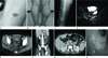
Fig. 1
An 18-year-old female patient diagnosed with Klippel-Trenaunay-Weber syndrome.
A. Cutaneous hemangioma on the right ankle.
B. Unilateral hypertrophy of soft tissue is noted in the right calf area.
C. Venography of the right leg obtained 14-year-old shows dilated superficial veins in the subcutaneous tissue.
D. Axial T2-weighted fat suppression magnetic resonance image obtained 14-year-old shows diffuse high signal intensity venous and lymphatic vascular malformations in the pelvic cavity, bilateral muscular compartment and subcutaneous tissue.
E, F. Axial (E) and coronal reconstruction CT (F) images show diffuse recto-sigmoid wall thickening (arrow), phleboliths and tubular enhanced vascular engorgement as a result of vascular malformation. Also note numerous venous malformations in the right buttock.
G. Axial CT scan shows multiple low density cystic masses in spleen without contrast enhancement, suggestive of lymphatic malformations of the spleen.
H. Colonoscopy shows vascular malformation involved the recto-sigmoid colon.
References
1. Cha SH, Romeo MA, Neutze JA. Visceral manifestations of Klippel-Trénaunay syndrome. Radiographics. 2005. 25:1694–1697.
2. Cheon SH, Lee SH, Park EB. Rectal involvement of Klippel-Trenaunay syndrome. J Korean Soc Coloproctol. 2009. 25:52–55.
3. Choi YJ, Jee SR, Park KS, Ryu CH, Seo HR, Ha SI, et al. [Involvement of splenic hemangioma and rectal varices in a patient with klippel: trenaunay syndrome]. Korean J Gastroenterol. 2011. 58:157–161.
4. Wilson CL, Song LM, Chua H, Ferrara M, Devine RM, Dozois RR, et al. Bleeding from cavernous angiomatosis of the rectum in Klippel-Trenaunay syndrome: report of three cases and literature review. Am J Gastroenterol. 2001. 96:2783–2788.
5. Kanterman RY, Witt PD, Hsieh PS, Picus D. Klippel-Trenaunay syndrome: imaging findings and percutaneous intervention. AJR Am J Roentgenol. 1996. 167:989–995.
6. Park MH, Kwon ST, Shin BS, Kim YM. An arteriovenous malformation in the suprapatellar fat pad of the knee associated with Klippel-Trenaunay-Weber syndrome: a case report. J Korean Radiol Soc. 2006. 54:27–31.
7. Lowe LH, Marchant TC, Rivard DC, Scherbel AJ. Vascular malformations: classification and terminology the radiologist needs to know. Semin Roentgenol. 2012. 47:106–117.
8. Yamazaki M, Kawamura Y, Ohka T, Katada S, Morita K, Nakagawa M, et al. Cavernous lymphangioma of the spleen in a patient with Klippel-Trenaunay-Weber syndrome. Intern Med. 1994. 33:574–577.
 XML Download