Abstract
Malignant mixed mullerian tumors (MMMTs) are a rare uterine tumor and contribute to approximately 1-3% of all corpus malignant tumors. MMMTs are usually in the uterine corpus, but can also arise from the uterine cervix, vagina, ovaries and fallofian tubes. MMMTs of the uterine cervix are extremely rare. MMMTs are highly malignant and tend to maintain a rapid growth and exhibit a high rate of recurrence. Therefore, the prognosis of patients diagnosed with these types of tumors is extremely poor. We report a rare case of a malignant mixed mullerian tumor arising from the uterine cervix and introduce CT and MRI findings. CT and magnetic resonance findings of the uterine cervical MMMT in our case show highly aggressive features, such as parametrial involvement, pelvic and paraaortic lymphadenopathy, and distant metastasis and high enhancement.
Figures and Tables
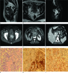
Fig. 1
Imaging and pathologic findings of 54-year-old woman with uterine cervical MMMT.
A. Sagittal T2 weighted image demonstrates large lobulated mass with Heterogenous slightly high signal intensity at the uterine cervix.
B. On axial T2 weighted image, low signal intensity cervical stoma is not visible. Right periureteric parametrial invasion and right hydroureter are also noted (arrow).
C. Gadolinium enhanced T1 weighted image shows strong enhancement of the mass and the epicenter of mass is located at the uterine cervix (arrow).
D. Contrast enhanced axial CT scan demonstrates highly enhanced uterine cervical mass.
E. On delayed scan, right hydronephrosis and metastatic paraaortic lymph nodes are identified.
F. Low attenuation metastatic nodule in liver is also noted (arrow).
G. On PAS staining (× 20), several cytoplasmic staining are identified (arrow).
H, I. The specimen shows high positivity on CEA (× 20) (H), and vimentin (× 20) (I) staining.
Note.-CEA = carcinoembryonic antigen, MMMT = malignant mixed mullerian tumor, PAS = Periodic Acid-Shiff

References
1. Sunwoo J, Cho IS, Jeon S, Bae DH, Shin YW, Kim CJ, et al. A case of malignant Mixed Mullerian Tumor (MMMT) of the uterine cervix. Korean J Obstet Gynecol. 2008. 51:350–354.
2. Sharma NK, Sorosky JI, Bender D, Fletcher MS, Sood AK. Malignant mixed mullerian tumor (MMMT) of the cervix. Gynecol Oncol. 2005. 97:442–445.
3. Grayson W, Taylor LF, Cooper K. Carcinosarcoma of the uterine cervix: a report of eight cases with immunohistochemical analysis and evaluation of human papillomavirus status. Am J Surg Pathol. 2001. 25:338–347.
4. Bharwani N, Newland A, Tunariu N, Babar S, Sahdev A, Rockall AG, et al. MRI appearances of uterine malignant mixed müllerian tumors. AJR Am J Roentgenol. 2010. 195:1268–1275.
5. Clement PB, Zubovits JT, Young RH, Scully RE. Malignant mullerian mixed tumors of the uterine cervix: a report of nine cases of a neoplasm with morphology often different from its counterpart in the corpus. Int J Gynecol Pathol. 1998. 17:211–222.
6. Teo SY, Babagbemi KT, Peters HE, Mortele KJ. Primary malignant mixed mullerian tumor of the uterus: findings on sonography, CT, and gadolinium-enhanced MRI. AJR Am J Roentgenol. 2008. 191:278–283.
7. Tanaka YO, Tsunoda H, Minami R, Yoshikawa H, Minami M. Carcinosarcoma of the uterus: MR findings. J Magn Reson Imaging. 2008. 28:434–439.
8. Seki H, Azumi R, Kimura M, Sakai K. Stromal invasion by carcinoma of the cervix: assessment with dynamic MR imaging. AJR Am J Roentgenol. 1997. 168:1579–1585.
9. Yamashita Y, Takahashi M, Sawada T, Miyazaki K, Okamura H. Carcinoma of the cervix: dynamic MR imaging. Radiology. 1992. 182:643–648.
10. Pannu HK, Corl FM, Fishman EK. CT evaluation of cervical cancer: spectrum of disease. Radiographics. 2001. 21:1155–1168.


 PDF
PDF ePub
ePub Citation
 Print
Print


 XML Download