

 PDF
PDF ePub
ePub Citation
 Print
Print


Abstract
We introduce two cases in which positron emission tomography (PET)/CT delineated viable malignant tissue from nonmalignant areas and guided us to successful biopsies when conventional CT failed to do so. According to our experience, PET/CT appears to be helpful in deciding the adequate site for biopsy in patients with lung cancer, owing to its capability to delineate malignant from nonmalignant areas, and also to reflect the areas with the most aggressive behaviors, especially in the era of the personalized cancer therapy.
Figures and Tables
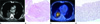 | Fig. 1A 53-year-old man with pleomorphic carcinoma.
A. Axial CT scan shows a homogeneously enhancing mass in the superior segment of the right lower lobe with right pleural effusion.
B. Photomicrograph from the first biopsy shows a core of fibrotic lung with necrosis (H&E, × 10).
C. PET-CT scan shows metabolic uptake, not in the entire tumor, but in deeper areas.
D. Photomicrograph from the rebiopsy shows malignant spindle cell tumor (H&E, × 40).
Note.-PET = positron emission tomography
|
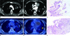 | Fig. 2A 44-year-old woman with adenocarcinoma.
A, B. Axial CT scans show multiple nodules with similar enhancements, including the largest one in the left upper lobe.
C. Photomicrograph from the first biopsy targeting the largest one of the left upper lobe shows only atypical columnar cells (H&E, × 40).
D, E. On the PET-CT scans, the lesion on the left upper lobes shows mild uptake; whereas the lesion in the left lower lobe shows strong uptake.
F. Photomicrograph from the rebiopsy targeting the lesion in the left lower lobe shows papillary adenocarcinoma with fibrovascular cores surrounded by malignant cells (H&E, × 40).
Note.-PET = positron emission tomography
|
References
1. Bomanji JB, Costa DC, Ell PJ. Clinical role of positron emission tomography in oncology. Lancet Oncol. 2001. 2:157–164.
2. Pauwels EK, Ribeiro MJ, Stoot JH, McCready VR, Bourguignon M, Mazière B. FDG accumulation and tumor biology. Nucl Med Biol. 1998. 25:317–322.
3. Travis WD, Brambilla E, Noguchi M, Nicholson AG, Geisinger KR, Yatabe Y, et al. International association for the study of lung cancer/american thoracic society/european respiratory society international multidisciplinary classification of lung adenocarcinoma. J Thorac Oncol. 2011. 6:244–285.
4. Prior JO, Stupp R, Christodoulou M, Letovanec I. Micro-papillary pattern in lung adenocarcinoma: aspect on 18F-fluorodeoxyglucose positron emission tomography/computed tomography imaging. Interact Cardiovasc Thorac Surg. 2010. 10:144–145.
5. Haruki T, Shomori K, Shiomi T, Taniguchi Y, Nakamura H, Ito H. The morphological diversity of small lung adenocarcinoma with mixed subtypes is associated with local invasiveness and prognosis. Eur J Cardiothorac Surg. 2011. 39:763–768.
6. Tatli S, Gerbaudo VH, Mamede M, Tuncali K, Shyn PB, Silverman SG. Abdominal masses sampled at PET/CT-guided percutaneous biopsy: initial experience with registration of prior PET/CT images. Radiology. 2010. 256:305–311.
7. Aquino SL, Halpern EF, Kuester LB, Fischman AJ. FDG-PET and CT features of non-small cell lung cancer based on tumor type. Int J Mol Med. 2007. 19:495–499.
8. Travis WD, Rekhtman N, Riley GJ, Geisinger KR, Asamura H, Brambilla E, et al. Pathologic diagnosis of advanced lung cancer based on small biopsies and cytology: a paradigm shift. J Thorac Oncol. 2010. 5:411–414.
9. Gonzalez-Angulo AM, Hennessy BT, Mills GB. Future of personalized medicine in oncology: a systems biology approach. J Clin Oncol. 2010. 28:2777–2783.
10. Rutman AM, Kuo MD. Radiogenomics: creating a link between molecular diagnostics and diagnostic imaging. Eur J Radiol. 2009. 70:232–241.
 XML Download