

 PDF
PDF ePub
ePub Citation
 Print
Print


Abstract
An enteric duplication is a relatively common congenital anomaly, which is rarely complicated by infection. We report the radiologic findings including ultrasound, barium enema and computed tomography (CT) of an infected colonic duplication that was confirmed by pathology. This case demonstrated a complex hypoechoic cystic mass with a thick wall and septa in the left lower quadrant of abdomen and increased the color flow on the Color Doppler ultrasonography. On CT images, the cystic mass contained multiple enhancing septa, infiltrated to the mesocolon and displaced the adjacent bowels. On exploration, a large cystic mass with an abscess attached to the mesocolic border adhering to the small bowel was found.
Figures and Tables
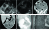
Fig. 1
A 8-day-old girl with infected colonic duplication.
A. Abdominal ultrasound shows a well-defined, lobulated, complex hypoechoic cystic mass with central echogenic septa (arrows) in left lower abdomen. Its wall is slightly thickened and contains hypoechoic internal septa.
B. Color Doppler ultrasound of the complex cystic lesion depicts increased color flow within the thickened septa (arrows).
C, D. The axial (C) and coronal reformatted images (D) of the contrast-enhanced abdominal CT scan shows a relatively well defined hypodense cystic mass (arrows) with heterogeneously mural enhancement and infiltrations in the mesocolon of the lower abdomen. This cystic mass surrounds the air containing sigmoid colon (arrowheads).
E. Barium enema reveals good contrast filling of the colon with segmental luminal narrowing of the sigmoid colon (arrows) due to external compression by cystic mass.
F. Photomicrograph (H&E, × 40) for a cross section of the cyst wall shows thickened and inflamed intestinal mucosa overlying the smooth muscle layers (arrows).
References
1. Ildstad ST, Tollerud DJ, Weiss RG, Ryan DP, McGowan MA, Martin LW. Duplications of the alimentary tract. Clinical characteristics, preferred treatment, and associated malformations. Ann Surg. 1988; 208:184–189.
2. Macpherson RI. Gastrointestinal tract duplications: clinical, pathologic, etiologic, and radiologic considerations. Radiographics. 1993; 13:1063–1080.
3. Jancelewicz T, Simko J, Lee H. Obstructing ileal duplication cyst infected with Salmonella in a 2-year-old boy: a case report and review of the literature. J Pediatr Surg. 2007; 42:E19–E21.
4. Yamauchi Y, Hoshino S, Yamashita Y, Funamoto S, Ishida K, Shirakusa T. Successful resection of an infected duodenal duplication cyst after percutaneous cyst drainage: report of a case. Surg Today. 2005; 35:586–589.
5. Lim GY, Im SA, Chung JH. Complicated duplication cysts on the ileum presenting with a mesenteric inflammatory mass. Pediatr Radiol. 2008; 38:467–470.
6. Caspi B, Schachter M, Lancet M. Infected duplication cyst of ileum masquerading as an adnexal abscess--ultrasonographic features. J Clin Ultrasound. 1989; 17:431–433.
7. Cheng G, Soboleski D, Daneman A, Poenaru D, Hurlbut D. Sonographic pitfalls in the diagnosis of enteric duplication cysts. AJR Am J Roentgenol. 2005; 184:521–525.
8. Barr LL, Hayden CK Jr, Stansberry SD, Swischuk LE. Enteric duplication cysts in children: are their ultrasonographic wall characteristics diagnostic? Pediatr Radiol. 1990; 20:326–328.
 XML Download