Abstract
Materials and Methods
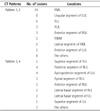
We retrospectively reviewed chest CT findings for 76 consecutive patients (21-84 years, average: 63 years; M : F = 30 : 46) who underwent an invasive diagnostic procedure under the suspicion of lung cancer and were pathologically diagnosed as pulmonary tuberculosis by bronchoscopic biopsy (n = 49), transthoracic needle biopsy (n = 17), and surgical resection (n = 10). We categorized the chest CT patterns of those lesions as follows: bronchial narrowing or obstruction without a central mass-like lesion (pattern 1), central mass-like lesion with distal atelectasis or obstructive pneumonia (pattern 2), peripheral nodule or mass including mass-like consolidation (pattern 3), and cavitary lesion (pattern 4). CT findings were reviewed with respect to the patterns and the locations of the lesions, parenchymal abnormalities adjacent to the lesions, the size, the border and pattern of enhancement for the peripheral nodule or mass and the thickness of the cavitary wall in the cavitary lesion. We also evaluated the abnormalities regarding the lymph node and pleura.
Results
Pattern 1 was the most common finding (n = 34), followed by pattern 3 (n = 23), pattern 2 (n = 11) and finally, pattern 4 (n = 8). The most frequently involving site in pattern 1 and 2 was the right middle lobe (n = 14/45). However, in pattern 3 and 4, the superior segment of right lower lobe (n = 5/31) was most frequently involved. III-defined small nodules and/or larger confluent nodules were found in the adjacent lung and at the other segment of the lung in 31 patients (40.8%). Enlarged lymph nodes were most commonly detected in the right paratracheal area (n = 9/18). Pleural effusion was demonstrated in 10 patients.
Figures and Tables
 | Fig. 1Pattern 1: Abrupt cutoff of bronchus without central mass-like lesion.
A. Chest radiograph in a 65-year-old woman shows ill-defined mass-like opacity in the right perihilar area.
B. CT scan at the carinal level shows abrupt cutoff (arrow) of anterior segmental bronchus of the right upper lobe with distal peribronchial consolidation.
C. Coronal reformatting CT scan shows enlarged lymph node in the right hilar area (arrow) without evidence of bronchial compression.
D. Follow-up chest radiograph after ten months of antituberculous chemotherapy shows marked contraction of the lesion.
|
 | Fig. 2Pattern 2: Bronchial obstruction with central mass-like lesion.
A. Lateral chest radiograph in a 68-year-old man shows atelectasis and consolidation in lower lung zones.
B-D. CT scans show concentric wall thickening of the right bronchus intermedius (arrow in B) and central mass-like lesion (arrow in C), which is obliterating the right lower lobar bronchial lumen, which has continuity to surrounding calcified lymph nodes (arrow in D) at a slightly upper level of C.
|
 | Fig. 3Pattern 3 (I): Heterogeneously enhancing peripheral mass.
A. Chest radiograph in a 35-year-old man shows approximately a 6 cm well defined mass-like opacity obscuring the diaphragmatic border in the right lower lung field and patchy and nodular increased opacity in the right mid lung field.
B. CT scan at the level of the inferior vena cava shows a heterogeneously enhancing mass containing multiple areas of necrotic low attenuation areas with peripheral rim enhancement and extension to the adjacent pleural space in the posterior aspect of the right basal lung. A small amount of pleural effusion is also noted (arrowheads).
C, D. CT scans at the level of the left atrium show another subpleural nodule (about 2 cm) with adjacent pleural thickening (arrowheads in C) and multiple small satellite nodules in the surrounding lung parenchyma (D, lung window setting).
|
 | Fig. 4Pattern 3 (II): Poorly enhancing peripheral nodule with necrotic lymphadenopathy.
A. Chest radiograph in a 37-year-old woman shows about a 1.5 cm nodule in the left upper lung field with multiple small nodules along the bronchovascular bundle in the surrounding lung field.
B. CT scan at the level of the aortic arch shows a poorly enhancing (mean HU: about 8) nodule in the apicoposterior segment of the left upper lobe.
C. CT scan with lung window setting shows small centrilobular nodules and linear branching opacities in the surrounding lung parenchyma.
D. CT scan at a slightly lower level shows an enlarged lymph node (arrow) containing low attenuation necrotic portion with peripheral rim-like enhancement in the aortopulmonary window area.
Note.-HU = Hounsfield unit
|
 | Fig. 5Pattern 4: Cavitary lesion with homogeneous low-attenuated wall.
A. Chest radiograph in a 60-year-old man shows about a 3.5 cm well-defined mass-like opacity in the right infrahilar area.
B, C. CT scans at the level of the left atrium show a cavitary mass (maximal thickness: about 12 mm) with a necrotic low attenuation wall (B, mean HU: about 4), speculated margin, and multiple small satellite nodules (C, lung window setting) in the superior segment of the right lower lobe.
Note.-HU = Hounsfield unit
|
References
1. Kuhlman JE, Deutsch JH, Fishman EK, Siegelman SS. CT features of thoracic mycobacterial disease. Radiographics. 1990. 10:413–431.
2. Im JG, Itoh H, Han MC. CT of pulmonary tuberculosis. Semin Ultrasound CT MR. 1995. 16:420–434.
3. Hatipoğlu ON, Osma E, Manisali M, Uçan ES, Balci P, Akkoçlu A, et al. High resolution computed tomographic findings in pulmonary tuberculosis. Thorax. 1996. 51:397–402.
4. Goo JM, Im JG. CT of tuberculosis and nontuberculous mycobacterial infections. Radiol Clin North Am. 2002. 40:73–87. viii
5. Andreu J, Cáceres J, Pallisa E, Martinez-Rodriguez M. Radiological manifestations of pulmonary tuberculosis. Eur J Radiol. 2004. 51:139–149.
6. Jeong YJ, Lee KS. Pulmonary tuberculosis: up-to-date imaging and management. AJR Am J Roentgenol. 2008. 191:834–844.
7. Lee JJ, Chong PY, Lin CB, Hsu AH, Lee CC. High resolution chest CT in patients with pulmonary tuberculosis: characteristic findings before and after antituberculous therapy. Eur J Radiol. 2008. 67:100–104.
8. Lee SW, Jang YS, Park CM, Kang HY, Koh WJ, Yim JJ, et al. The role of chest CT scanning in TB outbreak investigation. Chest. 2010. 137:1057–1064.
9. Schluger NW. CT scanning for evaluating contacts of TB patients: ready for prime time? Chest. 2010. 137:1011–1013.
10. Matthews JI, Matarese SL, Carpenter JL. Endobronchial tuberculosis simulating lung cancer. Chest. 1984. 86:642–644.
11. Maguire GP, Delorenzo LJ, Brown RB, Davidian MM. Endobronchial tuberculosis simulating bronchogenic carcinoma in a patient with the acquired immunodeficiency syndrome. Am J Med Sci. 1987. 294:42–44.
12. Lee KS, Kim YH, Kim WS, Hwang SH, Kim PN, Lee BH. Endobronchial tuberculosis: CT features. J Comput Assist Tomogr. 1991. 15:424–428.
13. Van den Brande P, Lambrechts M, Tack J, Demedts M. Endobronchial tuberculosis mimicking lung cancer in elderly patients. Respir Med. 1991. 85:107–109.
14. Oh YW, Kim JH, Chung HH, Kim KA. Differntiation between endobronchial tuberculosis and bronchogenic carcinoma associated with atelectasis or obstructive pneumonitis: CT evaluation. J Korean Radiol Soc. 1995. 33:537–543.
15. Lee JH, Chung HS. Bronchoscopic, radiologic and pulmonary function evaluation of endobronchial tuberculosis. Respirology. 2000. 5:411–417.
16. McAdams HP, Erasmus J, Winter JA. Radiologic manifestations of pulmonary tuberculosis. Radiol Clin North Am. 1995. 33:655–678.
17. Woodring JH, Vandiviere HM, Fried AM, Dillon ML, Williams TD, Melvin IG. Update: the radiographic features of pulmonary tuberculosis. AJR Am J Roentgenol. 1986. 146:497–506.
18. Goodwin RA, Des Prez RM. Apical localization of pulmonary tuberculosis, chronic pulmonary histoplasmosis, and progressive massive fibrosis of the lung. Chest. 1983. 83:801–805.
19. Hadlock FP, Park SK, Awe RJ, Rivera M. Unusual radiographic findings in adult pulmonary tuberculosis. AJR Am J Roentgenol. 1980. 134:1015–1018.
20. Armstrong P, Wilson AG, Dee P, Hansell DM. Imaging of diseases of the chest. 1995. 2nd ed. St. Louis: Mosby;77–96.
21. Itoh H, Tokunaga S, Asamoto H, Furuta M, Funamoto Y, Kitaichi M, et al. Radiologic-pathologic correlations of small lung nodules with special reference to peribronchiolar nodules. AJR Am J Roentgenol. 1978. 130:223–231.
22. Swensen SJ, Brown LR, Colby TV, Weaver AL, Midthun DE. Lung nodule enhancement at CT: prospective findings. Radiology. 1996. 201:447–455.
23. Yi CA, Lee KS, Kim EA, Han J, Kim H, Kwon OJ, et al. Solitary pulmonary nodules: dynamic enhanced multi-detector row CT study and comparison with vascular endothelial growth factor and microvessel density. Radiology. 2004. 233:191–199.
24. Honda O, Tsubamoto M, Inoue A, Johkoh T, Tomiyama N, Hamada S, et al. Pulmonary cavitary nodules on computed tomography: differentiation of malignancy and benignancy. J Comput Assist Tomogr. 2007. 31:943–949.
25. Pombo F, Rodríguez E, Mato J, Pérez-Fontán J, Rivera E, Valvuena L. Patterns of contrast enhancement of tuberculous lymph nodes demonstrated by computed tomography. Clin Radiol. 1992. 46:13–17.
26. Rho IG, Kook SH, Lee YR, Chin SB, Park YO, Park HW. CT findings of diffuse pleural diseases: differentiation of malignant diseases from tuberculosis. J Korean Radiol Soc. 1997. 36:619–625.
27. Kim YK, Kim HJ, Lee SW, Park SW, Lee SM, Cho KS, et al. Newly appearing tuberculous pulmonary masses during antituberculous treatment of tuberculous pleurisy: radiographic and CT findings. J Korean Radiol Soc. 2001. 45:597–603.


 PDF
PDF ePub
ePub Citation
 Print
Print


 XML Download