

 PDF
PDF ePub
ePub Citation
 Print
Print


Abstract
We report two cases of the aberrant vertebral artery, which originated from the part of the aortic arch about 2.5 cm distal to the left of the subclavian artery origin. The aberrant vertebral arteries were relatively hypoplastic. Herein, we review the previous reported cases in the literature and discuss embryologic basis and clinical implication of this variation.
Figures and Tables
Fig. 1
Images from a 72-year-old woman with severe headache. Aberrant right vertebral artery (arrows) arises directly from the proximal descending thoracic aorta, distal to the left subclavian artery. It courses behind the esophagus and the trachea (A) in front of the 3rd thoracic vertebral body, and ascends upward along the posterior mediastinum (B). Then, it enters the right transverse foramen of the 7th cervical vertebra (C).
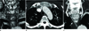
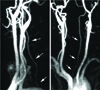
Fig. 2
Images from an 82-year-old man with transient right side weakness. Aberrant left vertebral artery (arrows) arises directly from the aortic arch about 2.6 cm distal to left subclavian artery. It ascends upward along the posterior mediastinum. It is relatively hypoplastic. Ascending aorta shows mild aneurysmal dilatation. The level of the entrance cannot be determined on magnetic resonance angiography.
References
1. Lemke AJ, Benndorf G, Liebig T, Felix R. Anomalous origin of the right vertebral artery: review of the literature and case report of right vertebral artery origin distal to the left subclavian artery. AJNR Am J Neuroradiol. 1999. 20:1318–1321.
2. Lasjaunias PL, Berenstein A, Brugge KGt. Lasjaunias PL, Berenstein A, Brugge KGt, editors. Spinal and spinal cord arteries and veins. Surgical neuroangiography. 2001. 2nd ed. Berlin, New York: Springer;82.
3. Schwarzacher SW, Krammer EB. Complex anomalies of the human aortic arch system: unique case with both vertebral arteries as additional branches of the aortic arch. Anat Rec. 1989. 225:246–250.
4. Karcaaltincaba M, Strottman J, Washington L. Multidetector-row CT angiographic findings in the bilateral aortic arch origin of the vertebral arteries. AJNR Am J Neuroradiol. 2003. 24:157.
5. Goray VB, Joshi AR, Garg A, Merchant S, Yadav B, Maheshwari P. Aortic arch variation: a unique case with anomalous origin of both vertebral arteries as additional branches of the aortic arch distal to left subclavian artery. AJNR Am J Neuroradiol. 2005. 26:93–95.
6. Satti SR, Cerniglia CA, Koenigsberg RA. Cervical vertebral artery variations: an anatomic study. AJNR Am J Neuroradiol. 2007. 28:976–980.
7. Son JS, Hong KB, Chung DC. Pseudocoarctation of the aorta associated with the anomalous origin of the left vertebral artery: a case report. Korean J Radiol. 2008. 9:283–285.
8. Hsu DP, Alexander AD, Gilkeson RC. Anomalous vertebral artery origins: the first and second reports of two variants. J Neurointerv Surg. 2010. 2:160–162.
9. Verin AL, Creuze N, Musset D. Multidetector CT scan findings of a right aberrant retroesophageal vertebral artery with an anomalous origin from a cervical aortic arch. Chest. 2010. 138:418–422.
10. Lacout A, Khalil A, Figl A, Liloku R, Marcy PY. Vertebral arteria lusoria: a life-threatening condition for oesophageal surgery. Surg Radiol Anat. 2012. 34:381–383.
 XML Download