

 PDF
PDF ePub
ePub Citation
 Print
Print


INTRODUCTION
Splenogonadal fusion is a rare congenital anomaly characterized as an abnormal fusion between the spleen and gonad. The diagnosis is almost always made when splenogonadal fusion presents as a scrotal mass, an incidental finding during surgery such as orchiopexy or inguinal hernioplasty (1). Moreover, this anomaly is most often misdiagnosed as an extratesticular tumor commonly requiring surgery such as orchiectomy, which could be avoided if there is a prior diagnosis (2). Multi-imaging modalities may reduce misdiagnoses and unnecessary surgery. In this view, we report an incidental case of splenogonadal fusion presenting as an extratesticular mass using multi-imaging modalities such as ultrasound, computed tomography (CT), magnetic resonance imaging (MRI), and scintigraphy. As far as we know, this is the first reported case of splenogonadal fusion in this country.
CASE REPORT
A 33-year-old man was referred to our hospital for evaluation of scrotal pain after trauma in June 2010. Upon physical examination, a 4-cm skin laceration of the left scrotum and mild scrotal swelling, as well as, a palpable left scrotal mass were noted. The man did not have any other noted abnormalities on physical examination. All laboratory investigations were normal.
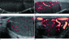
The patient underwent a scrotal ultrasound, which revealed a 3.3 × 3.0 cm oval-shaped relatively homogeneous isoechoic extratesticular mass at the left scrotum (Fig. 1). On grayscale and Doppler studies, multiple engorged vessels surrounding the mass were also noted. Bilateral testes and epididymides were normal in appearance without any traumatic complications.
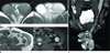
In the emergency room, the man complained of abdominal discomfort. To evaluate his abdominal injury, he underwent an unenhanced abdominal CT, which the presence of a left extratesticular mass with homogeneous density. A contrast-enhanced abdominal CT image obtained during the portal venous phase showed an elongated spleen extension to the left pelvic cavity and engorged splenic vessels which were connected with extratesticular mass in the left scrotum. The left extratesticular mass was homogeneously enhanced to a similar degree to that reof the spleen. A three-dimensional volume rendering CT image also demonstrated the elongated spleen, engorged splenic vessels driving together with the elongated spleen, and a left scrotal mass which was connected with the engorged splenic vessels. In addition, the engorged left gonadal vessels were identified (Fig. 2).
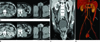
For further evaluation of the scrotal mass character, an abdominal MRI was performed, which revealed an extratesticular mass with similar signal intensity to the spleen on all sequences. Also, a well defined margin was identified between left testis and mass. The signal intensity of the testes and the left extratesticular mass was different on all sequences. The testes showed iso signal intensity on the T1-weighted image (WI) and high signal intensity on the T2WI, while the extratesticular mass showed iso signal intensity on both the T1 and T2WI, similar to the spleen (Fig. 3).
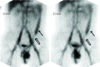
Planar scintigrams were performed after intravenous injection of 20 mCi of technetium-labelled red blood cells. A scan revealed a spleen elongated in shape with a tail-like projection 590at the left pelvic cavity and an oval shape with increased radioactivity in the left scrotum, which was connected with the spleen through band-like radioactivity (Fig. 4). Our patient showed continuous type splenogonadal fusion due to the connection between the spleen and gonad.
As all imaging findings were consistent with a splenogonadal fusion, and hence diagnosed as such. Because of a connection between the spleen and gonad, continuous type splenogonadal fusion was diagnosed. The patient received a dressing, suturing, and conservative management for the left scrotal skin laceration. But, any treatment for the splenogonadal fusion was not performed because the patient did not present with any symptoms related with the anomaly. He was discharged from the hospital for ambulatory follow-up.
DISCUSSION
Splenogonadal fusion is a rare congenital anomaly first reported in 1883 (3). Approximately 160 cases have been reported to date (2). Continuous and discontinuous types of splenogonadal fusion have been described. In the continuous form, a cord of the spleen or fibrous tissue extends from the normally sited spleen to the gonad. In the discontinuous form, no connection is found between the spleen and gonad (4). Both types appear with almost equal frequency (2).
Although splenogonadal fusion is found more commonly in males (256), the reported incidence of this anomaly in female patients may not reflect its true incidence based on the fact that an ovary cannot be explored as easily as testis. Most patients (82%) are younger than 30 years old; however, this anomaly may be diagnosed at any age (1-81 years) (1).
Splenogonadal fusion, especially the continuous type, has been associated with other congenital anomalies such as cryptorchidism, limb defects, micrognathia, anorectal abnormalities, spina bifida, cleft palate, facial muscle agenesis, peomelia, hypoglossia, polymicrohyria, Moebius syndrome, craniosynostosis, cardiac defects, thoracopagus, diaphragmatic hernia, abnormal lung fissure, hypoplastic lung, and hypospadias (1267). In our patient, no other abnormalities were observed.
The etiology of the malformation remains unclear. It most probably arises during the fifth through eighth week of embryonic life when the developing spleen is in intimate contact with the mesonephric-gonadal anlage. Sneath (8) suggested that inflammation of the peritoneal surfaces over the spleen and gonad can produce a partial fusion of the two organs.
Clinically, splenogonadal fusion occurs in 40% of cases as a paratesticular mass as in our patient. Occasionally, when the mass is closely attached to the testis, a differential diagnosis with paratesticular solid masses becomes very difficult (2).
Because of the rarity of this anomaly, it is seldom diagnosed preoperatively. Diagnosis is usually made during surgery for a paratesticular mass, inguinoscrotal hernia, or cryptorchidism. Imaging modalities for diagnosis are available if there is a clinical suspicion of splenogonadal fusion (16). Ultrasonic differences between the accessory spleen in the scrotum and normal testis are hard to find; however, ultrasonography is useful for the initial detection of a scrotal mass. The CT and MRI values for a diagnosis of splenogonadal fusion have the same density and signal intensity but also same degree of enhancement as an ectopic spleen in the scrotum compared with spleen. Some authors advocate the use of radionuclides with 99m Tc to confirm or rule out the presence of splenogonadal fusion (9). Among them, CT may be more useful because of its lower cost and shorter scan time.
The clinical importance of splenogonadal fusion is that it is most often misdiagnosed as an extratesticular tumor commonly requiring surgery such as an orchiectomy, which could be avoided if there is a prior correct diagnosis. In conclusion, a splenogonadal fusion should be considered as part of the differential diagnoses of an extratesticular mass.
 XML Download