

 PDF
PDF ePub
ePub Citation
 Print
Print


Abstract
Purpose
To compare radiation doses of dual-energy CT (DECT) to single-energy CT (SECT) by a phantom experiment, with the application of mean tube currents for abdomino-pelvic CT.
Materials and Methods
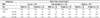
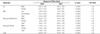
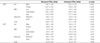
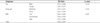
This study includes patients who were examined by contrast-enhanced CT for kidney evaluation. We divided the patients into six groups according to sex and body mass index. Each group consisted of five patients and a total of 30 patients were evaluated. We split the body parts (abdomen and pelvis), and calculated the mean tube current of each group as well as investigated the image noise. Applying the mean mAs from a CT scan, we measured the weighted CT dose index (CTDIw) of DECT and SECT. We compared the measured CTDIw to an estimated CTDI value displayed on the CT console. We also compared the radiation dose ratio of DECT to SECT (D/S ratio) for each subgroup. The radiation doses were compared by the student's t-test and analysis of variance.
Results
The difference of image noise between DECT and SECT was not statistically significant. Radiation dose of DECT was higher than SECT by about 21.6% (10.69 mGy, 8.79 mGy; p < 0.0001), and the measured CTDI of the DECT was significantly higher than the estimated CTDI by about 6% (p < 0.001). The D/S ratio was not significant between the six groups.
Figures and Tables

References
1. Flohr TG, Bruder H, Stierstorfer K, Petersilka M, Schmidt B, McCollough CH. Image reconstruction and image quality evaluation for a dual source CT scanner. Med Phys. 2008; 35:5882–5897.
2. Petersilka M, Bruder H, Krauss B, Stierstorfer K, Flohr TG. Technical principles of dual source CT. Eur J Radiol. 2008; 68:362–368.
3. Johnson TR, Krauss B, Sedlmair M, Grasruck M, Bruder H, Morhard D, et al. Material differentiation by dual energy CT: initial experience. Eur Radiol. 2007; 17:1510–1517.
4. Chae EJ, Seo JB, Goo HW, Kim N, Song KS, Lee SD, et al. Xenon ventilation CT with a dual-energy technique of dual-source CT: initial experience. Radiology. 2008; 248:615–624.
5. Goo HW, Chae EJ, Seo JB, Hong SJ. Xenon ventilation CT using a dual-source dual-energy technique: dynamic ventilation abnormality in a child with bronchial atresia. Pediatr Radiol. 2008; 38:1113–1116.
6. Nakayama Y, Awai K, Funama Y, Hatemura M, Imuta M, Nakaura T, et al. Abdominal CT with low tube voltage: preliminary observations about radiation dose, contrast enhancement, image quality, and noise. Radiology. 2005; 237:945–951.
7. Siegel MJ, Schmidt B, Bradley D, Suess C, Hildebolt C. Radiation dose and image quality in pediatric CT: effect of technical factors and phantom size and shape. Radiology. 2004; 233:515–522.
8. Graser A, Johnson TR, Bader M, Staehler M, Haseke N, Nikolaou K, et al. Dual energy CT characterization of urinary calculi: initial in vitro and clinical experience. Invest Radiol. 2008; 43:112–119.
9. Graser A, Johnson TR, Hecht EM, Becker CR, Leidecker C, Staehler M, et al. Dual-energy CT in patients suspected of having renal masses: can virtual nonenhanced images replace true nonenhanced images? Radiology. 2009; 252:433–440.
10. Ho LM, Yoshizumi TT, Hurwitz LM, Nelson RC, Marin D, Toncheva G, et al. Dual energy versus single energy MDCT: measurement of radiation dose using adult abdominal imaging protocols. Acad Radiol. 2009; 16:1400–1407.
11. McNitt-Gray MF. AAPM/RSNA Physics Tutorial for Residents: Topics in CT. Radiation dose in CT. Radiographics. 2002; 22:1541–1155.
12. Shope TB, Gagne RM, Johnson GC. A method for describing the doses delivered by transmission x-ray computed tomography. Med Phys. 1981; 8:488–495.
13. Bauhs JA, Vrieze TJ, Primak AN, Bruesewitz MR, McCollough CH. CT dosimetry: comparison of measurement techniques and devices. Radiographics. 2008; 28:245–253.
14. Perisinakis K, Damilakis J, Tzedakis A, Papadakis A, Theocharopoulos N, Gourtsoyiannis N. Determination of the weighted CT dose index in modern multi-detector CT scanners. Phys Med Biol. 2007; 52:6485–6495.
15. Goo HW. Individualized volume CT dose index determined by cross-sectional area and mean density of the body to achieve uniform image noise of contrast-enhanced pediatric chest CT obtained at variable kV levels and with combined tube current modulation. Pediatr Radiol. 2011; 41:839–847.
16. Schenzle JC, Sommer WH, Neumaier K, Michalski G, Lechel U, Nikolaou K, et al. Dual energy CT of the chest: how about the dose? Invest Radiol. 2010; 45:347–353.
17. Bauer RW, Kramer S, Renker M, Schell B, Larson MC, Beeres M, et al. Dose and image quality at CT pulmonary angiography-comparison of first and second generation dual-energy CT and 64-slice CT. Eur Radiol. 2011; 21:2139–2147.
18. Miyazaki O, Horiuchi T, Masaki H, Nosaka S, Miyasaka M, Tsutsumi Y, et al. Estimation of adaptive computed tomography dose index based on body weight in pediatric patients. Radiat Med. 2008; 26:98–103.
19. Nickoloff EL, Dutta AK, Lu ZF. Influence of phantom diameter, kVp and scan mode upon computed tomography dose index. Med Phys. 2003; 30:395–402.
 XML Download