

 PDF
PDF ePub
ePub Citation
 Print
Print


INTRODUCTION
Takayasu arteritis (TA) is a chronic inflammatory disease of unknown etiology primarily affecting the aorta and its main branches and the pulmonary arteries. The clinical features usually reflect limb or organ ischemia resulting from gradual stenosis of involved arteries. Takayasu arteritis often occurs in females during their reproductive years (1).
Sarcoidosis is a systemic granulomatous disease that commonly affects the hilar lymph nodes, lung parenchyma, skin, and almost any organ or tissue can be involved (2). Vasculitis may be found occasionally in patients with sarcoidosis. A few cases of concurrent sarcoidosis and TA have been reported (134). However, it has not been confirmed whether vasculitis is a true manifestation of the disease or an incidentally discovered abnormality. In this paper, we report a case of 55-year-old woman presenting with TA accompanied by sarcoidosis. This additional coincidence has allowed us to suspect a possible association between these two diseases.
CASE REPORT
A 55-year-old woman was referred with a 6-year history of dyspnea, and newly developed numbness and weakness of the left upper and both lower extremities for several months. The patient also complained of general weakness and dizziness. In addition, the patient has had recurrent uveitis for about one year. Family history was not significant. She did not smoke tobacco or drink alcohol and had no remarkable occupational exposures. A physical examination showed that the patient had a body temperature of 36.4℃, respiratory rate of 20 breaths per minute, and pulse rate of 76 beats per minute. Her blood pressure was 180/90 mm Hg in the right arm and immeasurable in the left arm. Bruits were observed over the bilateral carotid arteries, and a radial pulse could not be found in her left arm. Her white blood cell count and platelet count were 13,390 per mm3 and 227,000 per mm3, respectively Her erythrocyte sedimentation rate was slightly elevated at 32 mm/hour and her serum complement levels were normal. Her serum angiotensin-converting enzyme (ACE) level was elevated at 84.7 U/mL. Tests for antineutrophil cytoplasmic antibodies were negative. Other negative serological studies included antinuclear antibody, anti-DNA antibody, rheumatoid factor, cryoglobulin, anti-human immunodeficiency virus, syphilis, and hepatitis C.
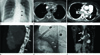
A chest radiograph demonstrated widening of the mediastinal silhouette and severe calcification of the descending aorta (Fig. 1A). A contrast-enhanced computed tomography (CT) of the chest using a 16-multi-detector CT (LightSpeed, GE Healthcare, Milwaukee, WI, USA) was performed. The CT images revealed bilateral enlarged mediastinal and hilar lymph nodes without necrosis, and extensive calcification with narrowing of the aorta and major its branches (Fig. 1B-D). Selective arteriography showed near total occlusion of the left subclavian artery with collateral vessels (Fig. 1E). 18-F-fluorodeoxyglucose positron emission tomography/CT (Biograph Duo, Siemens, Germany) revealed increased uptake in the bilateral mediastinal lymph nodes with maximum SUV 7.5 (Fig. 1F).
From all of these findings, differential diagnosis included Takayasu's arteritis combined with sarcoidosis, lymphoma or metastatic adenopathy. An excisional biopsy of the right lower paratracheal lymph nodes was performed through a right thoracotomy. Histologic examination showed granulomatous inflammation consistent with sarcoidosis. The result of a tissue culture was negative for tuberculosis. The patient began a regimen of prednisolone at 32 mg/day. Two weeks after the prednisolone treatment, the patient's blood pressure was 166/89 mm Hg in the right arm and 98/68 mm Hg in the left arm. After 3 months of treatment, her serum ACE level dropped to normal (32.2 U/mL). After 6 months of treatment, prednisolone treatment was gradually tapered to 8 mg/day and her symptoms improved.
DISCUSSION
According to the classification criteria of the 1990 American College of Rheumatology, Takayasu's arteritis can be classified if 3 out of the following 6 criteria are fulfilled: 1) age of 40 years or younger at disease onset, 2) claudication of the extremities, 3) decreased brachial artery pulse, 4) blood pressure difference between both arms of more than 10 mm Hg, 5) bruits over subclavian arteries or aorta, and 6) arteriographic abnormalities (5). Our patient fulfilled 5 of the criteria, except for age, so that late-onset of Takayasu's arteritis is more likely to be the diagnosis for this patient. A rare and interesting manifestation of this patient is the coexistence of Takayasu's arteritis with mediastinal lymphadenopathy, which was confirmed with sarcoidosis on pathology. According to Fernandes et al. (6), sarcoidosis may be complicated by systemic vasculitis that can affect small- to large-caliber vessels, and reported that among patients with large vessel disease, African Americans and Asians appear to be overrepresented in the United States. To the best of our knowledge, only six cases of Takayasu's arteritis associated with sarcoidosis were reported in English literature (134). However, focus has been on the clinical symptoms and etiology, with little emphasis on the imaging appearance.
The scarcity of reported cases in the literature suggests that the association between sarcoidosis and Takayasu's arteritis may be coincidental. However, considering the relatively low prevalence of sarcoidosis and TA (sarcoidosis: 1 to 4/million persons; TA: 1 to 2.6/million persons in western hemisphere and up to 30/million in Japanese annually), the concurrence of Takayasu's arteritis with sarcoidosis exceeds the expected incidence and raises the possibility that these conditions may be pathogenically related. In fact, in a review of 60 patients with TA, Kerr et al. (1) found two cases of sarcoidosis associated with TA and Fernandes et al. (6) suggested that TA or other forms of vasculitis and sarcoidosis may be related diseases (7). Reports of Crohn's disease, another granulomatous disorder, with TA further support the possibility of a common basis of granulomatous disorders and TA (1). In a review of the concurrence of sarcoidosis and aortitis, Weiler et al. (3) described common features in almost all reported cases: 1) sarcoidosis precedes vasculitis with a time lag of several years between diagnoses; 2) the aorta and/or its major branches are affected; 3) 50% of patients have uveitis, which is much less common in frank sarcoidosis; 4) all patients respond to glucocorticoid treatment.
In the current report, diagnosis of sarcoidosis and TA were apparently made at the same time, so that the time lag between these two diseases was not revealed. We assume that the reason for delayed diagnosis of stage 1 sarcoidosis in this case is due to silent mediastinal adenopathy.
According to Park et al. (8), the presence of a uniformly thickened, concentric, high attenuation wall in the aorta or the presence of extensive full-thickness calcification in a relatively young woman strongly suggests the possibility of Takayasu arteritis. In our case of a middle-aged woman, unusually extensive calcification along the descending aorta seemed to be related to the late onset of TA. It has been suggested that the thickened wall of calcification in TA tends to be linear in shape and present in the aortic arch and descending aorta, sparing the ascending aorta (9), which was also found in our case.
Like in our case of TA with bilateral mediastinal lymphadenopathy, other possibilities of mediastinal malignancy including lymphoma or metastatic adenopathy could not be excluded only by imaging features in a middle-aged or elderly woman. According to the probable relationship between TA and sarcoidosis presented as sporadic reports, however, sarcoidosis should be another diagnosis of choice for mediastinal lymphadenopathy, even if the patients of TA were old enough for a malignancy to be considered. In conclusion, we report a case of concurrence of TA and sarcoidosis showing bulky mediastinal widening and extensive aortic calcification with luminal narrowing on the chest radiograph in a middle-aged woman.
 XML Download