

 PDF
PDF ePub
ePub Citation
 Print
Print


INTRODUCTION
Marchiafava-Bignami Disease (MBD) is a disease observed in chronic alcoholic patients which results from the symmetric demyelination of the corpus callosum (1). The clinical diagnosis of MBD can be difficult. Recent magnetic resonance (MR) imaging including diffusion-weighted imaging (DWI) have been helpful in analyzing the distribution of lesions as well as establishing the diagnosis. We report here on a case of MBD with symmetric involvement of the corpus callosum, white matter, corticospinal tract, internal capsule, and middle cerebellar peduncle.
CASE REPORT
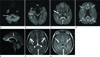
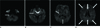
A 49-year-old man was referred to the emergency department with dizziness, trembling hands and weakness in both legs for a day. The patient had been an alcoholic with a daily intake of alcohol. DWI obtained at admission showed symmetrical hyperintense lesions involving the corpus callosum, white matter, corticospinal tract, internal capsule, and middle cerebellar peduncle (Fig. 1). Fluid-attenuated inversion recovery (FLAIR) and T2 weighted images (T2WI) showed subtle high signal intensity and apparent diffusion coefficient mapping, which showed relatively high hypointensity in those lesions. No further enhancement of the lesions was observed after gadolinium. On the basis of the clinical history and imaging findings, MBD was diagnosed. The patient was admitted and given intravenous thiamine, which resulted in an almost complete remission of the symptoms related to the disease. Follow-up MR imaging performed 11 months after the onset showed the disappearance of signal-intensity abnormalities on FLAIR, T2WI, and DWI (Fig. 2).
DISCUSSION
MBD is a rare form of toxic demyelination and necrosis of the corpus callosum associated with chronic alcohol consumption. This disease is occasionally observed in non-alcoholic patients as well. It is generally accepted that the cause of the disease is mainly due to the deficiency of vitamin B complex (159). Most patients are male, between 40 and 60 years of age, and have a history of chronic alcoholism with poor oral intake (12).
The main pathologic change associated with MBD is a degeneration of the corpus callosum with different degrees of damage, from demyelination with preservation of axons, to 462necrosis. The genu is most frequently involved, but the degeneration can extend to the entire corpus callosum (56). Other white matter tracts such as the anterior and posterior commisures, corticospinal tracts, hemispheric white matter, and middle cerebellar peduncle may also be involved (2). Selective involvement of the entire length of the corpus callosum and chronic alcoholism, other clinical features, and MRI findings support the diagnosis (1). MR imaging is the best technique
for evaluating MBD lesions (3). Characteristic MR imaging findings are symmetric lesions on the corpus callosum; but, the lesions may be also found in the hemispheric white matter, cortex, middle cerebellar peduncles, and internal capsules (4). In the acute phase, the affected areas are hypointense on T1WI and hyperintense on T2WI, due to edematous change with or without demyelination. However, these lesions do not have mass effect and may show peripheral contrast enhancement during the acute stage (2). At 30 hrs after initial imaging, hyperintense signals on DWI appeared which was in contrast to the initial image where the signals did not appear. These findings suggest that the initial change in the corpus callosum was partly attributed to vasogenic edema and that the lesion was then converted into cytotoxic edema. The necrotic lesions in the corpus callosum in chronic stage confirmed the cytotoxic process (7).
Acute MBD is a rare toxic disease, but typical history, clinical manifestation, and MR findings with DWI could facilitate its diagnosis.
 XML Download