

 PDF
PDF ePub
ePub Citation
 Print
Print


INTRODUCTION
Regarding the lumbar spine, common iliac vessels have been mentioned only for injury as one of the perioperative complications of the lumbar spine in previously published reports (123). There have been no reports about the imaging or surgical findings of intradiscal herniation of iliac vessels. We report on a case of the bifurcations of the common iliac vessels that herniated into the L4-L5 disc.
CASE REPORT
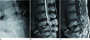
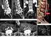
A 65-year-old woman presented with aggravated chronic pain and numbness in both lower extremities, as well as lower back pain for several decades. She had no previous history of trauma or lumbar spine surgery; a lateral plain radiograph of the lumbar spine revealed isthmic spondylolisthesis with severe disc degeneration at L4-L5, as well as severe disc degeneration with retrolisthesis at L5-S1. An anterior wedged deformity of the L5 body was noted (Fig. 1A), as well as segmental instability at L4-L5 on flexion-extension radiographs (not shown). Magnetic resonance imaging of the lumbar spine demonstrated herniation of iliac vessels with surrounding fat into the L4-L5 disc (Fig. 1B, C). Central spinal canal narrowing at the level of L5 was shown (Fig. 1B). Computed tomography (CT) angiography of the lumbar spine demonstrated the bifurcation of the right common iliac artery and common iliac veins sandwiched between the anterior bodies of L4 and L5 (Fig. 2A-F).
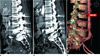
Disc degeneration at L4-L5 and L5-S1 with sclerosis of bodies, tiny erosions or cysts at endplates, vacuum disc phenomena, and disc height loss as well as anterior wedged deformity of L5 body (Fig. 2G), were noted. A chronic compression fracture of the L5 body was suggested, despite the fact that the patient had no history of trauma. Consequently, the patient underwent posterior decompression and posterolateral fusion at L4-S1 for isthmic spondylolisthesis and severe disc degeneration with segmental instability as well as central spinal canal narrowing (not shown). After that, the patient underwent revision posterior fusion with instrumentation and an auto-iliac bone graft at L2-S1 because of continuing pain, as well as loosening around the screws on postoperative CT with signs of infection (Fig. 3). There was no more atypical location of the iliac vessels between the bodies of L4 and L5 on postoperative CT angiography of the lumbar spine. The patient required treatment for pain reduction with occasional milder attacks at a year and a half follow-up.
DISCUSSION
The atypical location of iliac vessels between vertebral bodies may be formed through defects of the anterior longitudinal ligament and annulus fibrosus or with severe laxity of the anterior longitudinal ligament. The anterior longitudinal ligament is a strong ligament running down the anterior surface of the spine. The annulus fibrosus is an outer part of the intervertebral disc surrounding the nucleus pulposus. The annulus fibrosus is a strong ring of elastic collagen, in which the collagen fibers are arranged obliquely in alternating directions and in layers (lamellae), and is comprised of 15 to 25 lamellae. Collagen fibers continue from the annulus into the adjacent tissues, which attach this structure to each vertebral body at its rim, and furthermore to the anterior and posterior longitudinal ligaments, as well as to the hyaline cartilage endplates superiorly and inferiorly (45). The anterior longitudinal ligament and the annulus fibrosus have a self-sealing effect be-cause of their strong and elastic nature (67).
In our case, chronic isthmic spondylolisthesis with segmental instability, anterior wedged deformity of the L5 body, and retrolisthesis of L5 on S1 might cause a defect or severe laxity of the anterior longitudinal ligament at the level of the L4-L5 disc and L5, resulting in intradiscal herniation of the iliac vessels. A chronic compression fracture was suggested for the anterior wedged deformity of the L5 body, although the patient had no history of trauma. Furthermore, the sequelae of infectious spondylitis could not be ruled out, even with a lower probability. We thought that there was no possibility of the congenital posterior hemivertebra on the basis of no anomaly of the L4 body and L4-L5 disc.
In the patient with atypical positioning of the iliac vessels between the vertebral bodies and lumbar segmental instability, there might be a higher risk of injury of the iliac vessels by trauma or strenuous exercise; therefore, recognition of these conditions and attention may be needed. The decision between medical treatment and surgery may be influenced by these conditions as well. The radiologists should be concerned with the retroperitoneal structures just anterior to the spine, as well as the spine, disc, and spinal canal on images of the spine so as to provide information to clinicians.
In conclusion, this is the first case description of iliac vessels herniating into the L4-L5 disc associated with isthmic spondylolisthesis and segmental instability.
 XML Download