

 PDF
PDF ePub
ePub Citation
 Print
Print


Abstract
Purpose
To prospectively evaluate the usefulness of the fluoroscopically-guided posterior approach compared with the anterior approach for shoulder magnetic resonance (MR) arthrography.
Materials and Methods
Institutional review board approval and informed consent were obtained. Among 60 shoulder MR arthrographies performed on 59 patients with symptomatic shoulders, an intra-articular injection was performed (30 cases using the anterior approach and 30 using the posterior approach). Procedure-related pain was assessed by using a 5 score visual analogue scale (VAS). Depth of the puncture and standardized depth of puncture by body mass index (BMI) were recorded. The contrast leakage along the course of the puncture was evaluated by reviewing the MR. The statistical analyses included the Mann-Whitney U and Kruskal-Wallis test.
Results
There was no significant difference in VAS scores between the anterior and posterior groups (1.77 ± 1.10 vs. 1.80 ± 0.96). Depth of puncture and standardized depth of puncture by BMI were significantly shorter in the posterior group than those in the anterior group (4.4 ± 0.8 cm and 1.8 ± 0.3 cm vs. 6.6 ± 0.9 cm and 2.8 ± 0.4 cm, p < 0.001), respectively. The incidence of contrast leakage was more frequent in the posterior group (p = 0.003).
Figures and Tables
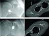 | Fig. 1Position and puncture site of anterior and position approach in shoulder magnetic resonance arthrography. The puncture site of the anterior approach (A) is targeted on the inferior portion of the joint space and the punctural tract (arrow and broken line) on CT (B) is penetrated through pectoralis major and minor and subscapularis muscles. The puncture site of the posterior approach (C) is targed on the posteromedial portion of the humeral head just lateral to joint space and the punctural tract (arrow and broken line) on CT (D) is through deltoid and infraspinatus muscles.
|
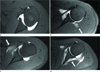 | Fig. 2Contrast leakage on shoulder magnetic resonance arthrography with fat suppressed T1-weighted images with axial plane.
A. Grade 0 presented no contrast leakage.
B. Grade 1 is defined as contrast leakage along the puncture tract which is localized within the only one muscle bundle.
C. Grade 2 is defined as contrast leakage beyond a muscle bundle extending to the intermuscular fascial plane or other adjacent muscle bundles.
D. Grade 3 shows separation of the muscle from scapula owing to the deep penetration of the contrast.
|
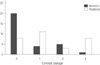
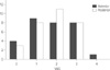 | Fig. 3Frequency of visual analogue scale (VAS) score on both anterior and posterior approach for magnetic resonance arthrography. VAS of the anterior approach group shows grade 1 is most frequent, but VAS of the posterior approach group is most frequent on grade 2. There is not statistically different between two groups.
|
References
1. Flannigan B, Kursunoglu-Brahme S, Snyder S, Karzel R, Del Pizzo W, Resnick D. MR arthrography of the shoulder: comparison with conventional MR imaging. AJR Am J Roentgenol. 1990; 155:829–832.
2. Chandnani VP, Yeager TD, DeBerardino T, Christensen K, Gagliardi JA, Heitz DR, et al. Glenoid labral tears: prospective evaluation with MRI imaging, MR arthrography, and CT arthrography. AJR Am J Roentgenol. 1993; 161:1229–1235.
3. Oberholzer J. Die Arthropneumoradiographie bei Habitueller Shulterluxation. Röntgen praxis. 1933; 5:589–590.
4. Schneider R, Ghelman B, Kaye JJ. A simplified injection technique for shoulder arthrography. Radiology. 1975; 114:738–739.
5. Petersilge CA, Lewin JS, Duerk JL, Hatem SF. MR arthrography of the shoulder: rethinking traditional imaging procedures to meet the technical requirements of MR imaging guidance. AJR Am J Roentgenol. 1997; 169:1453–1457.
6. Farmer KD, Hughes PM. MR arthrography of the shoulder: fluoroscopically guided technique using a posterior approach. AJR Am J Roentgenol. 2002; 178:433–434.
7. Chung CB, Dwek JR, Feng S, Resnick D. MR arthrography of the glenohumeral joint: a tailored approach. AJR Am J Roentgenol. 2001; 177:217–219.
8. Jacobson JA, Lin J, Jamadar DA, Hayes CW. Aids to successful shoulder arthrography performed with a fluoroscopically guided anterior approach. Radiographics. 2003; 23:373–378. discussion 379.
9. Dépelteau H, Bureau NJ, Cardinal E, Aubin B, Brassard P. Arthrography of the shoulder: a simple fluoroscopically guided approach for targeting the rotator cuff interval. AJR Am J Roentgenol. 2004; 182:329–332.
10. Zwar RB, Read JW, Noakes JB. Sonographically guided glenohumeral joint injection. AJR Am J Roentgenol. 2004; 183:48–50.
11. Gokalp G, Dusak A, Yazici Z. Efficacy of ultrasonography-guided shoulder MR arthrography using a posterior approach. Skeletal Radiol. 2010; 39:575–579.
12. Redondo MV, Berná-Serna JD, Campos PA, Reus M, Martínez F, Campos M, et al. MR arthrography of the shoulder using an anterior approach: optimal injection site. AJR Am J Roentgenol. 2008; 191:1397–1400.
13. Binkert CA, Zanetti M, Hodler J. Patient's assessment of discomfort during MR arthrography of the shoulder. Radiology. 2001; 221:775–778.
14. Saupe N, Zanetti M, Pfirrmann CW, Wels T, Schwenke C, Hodler J. Pain and other side effects after MR arthrography: prospective evaluation in 1085 patients. Radiology. 2009; 250:830–838.
15. Steurer-Dober I, Rufibach K, Hodler J, Saupe N, Zanetti M, Fucentese SF, et al. Do patients with structural abnormalities of the shoulder experience pain after MR arthrography of the shoulder? Radiology. 2010; 256:870–878.
16. Emig EW, Schweitzer ME, Karasick D, Lubowitz J. Adhesive capsulitis of the shoulder: MR diagnosis. AJR Am J Roentgenol. 1995; 164:1457–1459.
17. Seeger LL, Gold RH, Bassett LW. Shoulder instability: evaluation with MR imaging. Radiology. 1988; 168:695–697.
 XML Download