

 PDF
PDF ePub
ePub Citation
 Print
Print


INTRODUCTION
Lower extremity deep venous thrombosis (DVT) is a serious medical condition that can result in death or major disability due to pulmonary embolism and post-thrombotic syndrome (PTS). Although anticoagulation (heparin followed by oral anticoagulation) is currently considered the standard of care for the prevention of pulmonary embolism and recurrent DVT, this form of therapy does not protect the patient from the manifestations of PTS, which can appear months to years after the acute episode of DVT. Early thrombus removal is a logical approach to improve the long-term outcome of DVT. Two goals may be achieved by early thrombus removal: relief of venous outflow obstruction and preservation of valve function, both of which are established determinants of PTS.
Treatment strategies of DVT have included systemic thrombolytic therapy, surgical thrombectomy, and catheter-directed thrombolysis. Among them, catheter-directed thrombolysis is an attractive method, because it can potentially help achieve restoration of the lumen and removal of the thrombus lining the venous valves (123). However, the risk of major bleeding complications during venous thrombolysis has varied significantly in published studies and represents a significant impediment to widespread acceptance of catheter-directed thrombolysis for the treatment of the DVT (123). Several studies have demonstrated that mechanical thrombectomy (MT) devices can remove venous thrombus quickly and effectively without the bleeding risks associated with pharmacologic thrombolysis (45).
The purpose of this study was to evaluate the safety and efficacy of short-term catheter-directed thrombolysis using low-dose urokinase (UK) using an adjunctive method.
MATERIALS AND METHODS
Patients
This study was approved by the institutional review board. The radiology information systems database was searched for patients who underwent catheter-directed thrombolysis for DVT between January 2006 and April 2010. Patients were included in this retrospective analysis if they had documented sonographic, computed tomographic, and/or venographic confirmation of deep vein thrombosis. Patients were excluded from this retrospective analysis if UK was administered continuously overnight (> 8 hours). Inclusion criteria for this study were applied to low-dose UK as either a single intravenous bolus or as a continuous infusion for less than 8 hours. Consequently, eighty-nine patients (46 women and 43 men; age range, 16-86 years; mean 58.1 years) were evaluated. All patients did not have a contraindication for the use of anticoagulation, contrast media, or thrombolytic agents. Contraindications for thrombolytic agents included active internal bleeding, recent cerebrovascular accident, allergy to thrombolytic agents, recent major surgery, recent serious gastrointestinal bleeding, recent serious trauma, and coagulopathy. Initial symptoms, duration of symptoms, predisposing factors, and thrombus location were recorded for each treated extremity.
Endovascular Procedure
The physician who performed the procedure selected the catheterization technique for thrombolysis. The venous access sites included the internal jugular vein, the common femoral vein, and the popliteal vein. Over time, the ipsilateral popliteal venous approach became the access site of choice. In a typical case, with the patient prone on the angiographic table, the popliteal vein was accessed under ultrasonographic guidance with a small gauge needle. Most commonly, a 5-F sheath was inserted and all subsequent catheter and wire exchanges were performed through it. Infusion was performed by using the end hole catheter and/or a multiple-side-hole infusion catheter (Multi-sideport catheter infusion set, Cook, Bloomington, IN, USA). Before thrombolytic therapy, venography was once again performed to confirm the position and evaluate the inferior vena cava (IVC) thrombus. Prophylactic inferior vena cava filter placement was not routinely performed; the catheter was placed two thirds of the distance into the thrombosed venous segment and urokinase (Urokinase, Green Cross, Seoul, Korea) was administered either as a single bolus (100,000-150,000 IU) or as a continuous infusion (100,000-150,000 IU/h in split doses). Patients were systemically anticoagulated with a 5,000 IU bolus of intravenous heparin.
After catheter-directed thrombolysis, aspiration thrombectomy in 67 (75%) patients and/or mechanical thrombectomy in 30 (34%) patients were performed in an attempt to clear large volumes of softening thrombi. Aspiration thrombectomy was performed in a to-and-fro fashion using an 8-10 F guiding catheter (Envoy catheter, Cordis, Miami, FL, USA), while maintaining negative pressure with a 50 mL syringe. To prevent body volume loss during aspiration thrombectomy, the same amount of heparinized saline was infused manually via the sheath side arm. A mechanical thrombectomy was performed using a Trerotola Percutaneous Thrombectomy Device (Arrow International, Reading, PA, USA) to fragment the organized thrombus.
The status of lysis was monitored at the venography at 30 minutes intervals. If only partial lysis was achieved, the infusion catheter was repositioned within the residual thrombus and the infusion was continued. The start and end times of thrombolysis, as well as the concentration and total amount of UK administered, were recorded. If UK was applied as a single bolus injection, the infusion time was less than 10 seconds. In patients with persistent occlusion of the iliac segment but a patent femoro-popliteal segment, the iliac vein was recanalized by means of angioplasty only or followed by stent deployment. Initial dilatation for reconstruction of the iliac vein was achieved with a 8-12-mm-diameter balloon catheter (Cook, Bloomington, IN, USA). Stents were placed over a guide wire into the iliac vein and were typically between 10 to 14 mm in diameter and 6 to 8 cm long. Zilver stents (Cook, Bloomington, IN, USA; n = 25) and Smart stents (Cordis, Miami, FL, USA; n = 37) were commonly used to reconstruct the iliac vein. The deployed stents were fully dilated with the appropriate diameter angioplasty balloon catheter.
After endovascular therapy, 6 months of anticoagulation therapy with warfarin sodium was initiated and adjusted to maintain the international normalized ratio in the range of 2.0-3.0. Patients were subsequently fitted for a knee- or thigh-high venous compression hose.
Outcome Variables
The patients' medical records, radiology reports, procedural data, and venograms were reviewed. Immediate venous patency was evaluated in terms of technical success and clinical success. Assessment of immediate technical success was retrospectively performed by one radiologist on examination of the initial venographic images and the images obtained after completion of the interventional procedure. Technical success was defined as either the complete thrombolysis of clot, technical restoration of normal venous blood flow, and less than 30% remaining luminal narrowing; or partial thrombolysis of clot that allows adjunctive methods (such as angioplasty, stent placement, or thrombectomy) to be used for the successful restoration of patency and flow (6789). Immediate clinical success was defined as the presence of technical success in conjunction with considerable improvement in lower extremity swelling and/or pain before hospital discharge.
Late venous patency was evaluated in terms of primary patency rate and/or clinical success. Because objective imaging follow-up to assess for recurrent DVT and valvular dysfunction was not routinely obtained, late primary patency rate was evaluated in patients who underwent follow-up computed tomography angiography (CTA) or Doppler sonography after 6 months and 12 months at the end of the catheter-directed thrombolysis. Primary patency was defined as the time from the intervention, to the first occurrence of either thrombosis of the treated segment or to an intervention for maintaining patency (6). For patients who were lost to a more recent follow-up, the follow-up interval was defined as the time between the venous intervention and the last time point at which information about the clinical limb status was clearly documented in the medical record. Clinical success was graded as completely improved (asymptomatic), partially improved (moderately improved), unchanged, or worse.
Major bleeding was defined as intracranial bleeding, bleeding resulting in death, transfusion, surgery, or cessation of thrombolytic therapy, according to Society of Interventional Radiology reporting standards (10).
Data Analysis and Statistics
The technical success rates, clinical success rates, primary patency rates, and incidences of major bleeding were expressed as percentages. The mean thrombolytic infusion times, total thrombolytic agent doses, and their standard deviations were calculated and the primary patency rate calculated at 6 and 12 months, respectively.
RESULTS
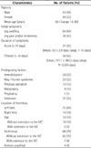
All patients were symptomatic with one or more of the following: leg swelling, leg pain and/or tenderness (Table 1). In 31 (35%) patients, acute DVT (defined as symptom duration of 10 days or less) was treated. In 58 (65%) patients, chronic DVT (defined as symptom duration of greater than 10 days) was treated. At the time of treatment, nine (10%) patients had a known malignancy and one (1%) patient was pregnant. The location of the treated thrombosed venous segments was the left limb in 75 (84%) patients and the right limb in 14 (16%) patients. As determined at venography, iliofemoral DVT was present in 69 (78%) patients, in which 7 (8%) where the thrombus extended into the IVC. Isolated femoro-popliteal DVT was found in 4 (4%) patients, whereas isolated iliac DVT was found in 16 (18%) patients.
Thirty-six (40%) patients were given a single bolus injection of UK (range, 4-14 × 104 IU, mean dose, 4.89 ± 2.51 × 10⁴ IU) and 53 (60%) patients had a continuous infusion of UK (range, 12-80, mean dose, 33.73 ± 16.42 × 10⁴ IU) for a mean of 168 minutes (range, 30-420 minutes) (Table 2). After catheter-directed thrombolysis, aspiration thrombectomy with or without mechanical thrombectomy was performed in 67 (75%) patients. Subsequent angioplasty and stent placement was performed in 83 (93%) patients and 65 (73%) patients, respectively. The rate of aspiration thrombectomy with or without mechanical thrombectomy was higher in acute DVT patients (97%) than in chronic DVT patients (64%). Balloon angioplasty was performed in 30 (97%) acute DVT patients and stents were placed in 25 (81%) acute DVT patients. Balloon angioplasty was performed in 53 (91%) chronic DVT patients and stents were placed in 40 (69%) chronic DVT patients.
Continuous UK infusion was performed in 15 (48%) acute DVT patients and 38 (66%) chronic DVT patients (Table 2). In acute DVT patients, the mean amount of UK delivered in a single dose and continuous infusion was 4.8 ± 2.5 × 10⁴ IU and 29.3 ± 12.6 × 10⁴ IU, respectively, in chronic DVT patients, the mean amount of UK delivered in a single dose and continuous infusion was 4.7 ± 2.2 × 10⁴ IU and 36.4 ± 18.2 × 10⁴ IU, respectively. The mean amount of continous UK infusion was higher in chronic DVT patients than in acute DVT patients. Also, both mean UK infusion time and total procedure time were higher in chronic DVT patients than in acute DVT patients.
Immediate technical success was achieved in all patients by completion venography. Clinical success, defined as a decrease in swelling, pain and/or swelling of the affected extremity within hospital discharge, was displayed in 80 (90%) patients. There was no major complication during or after the procedure.
Late anatomic success was evaluated in patients who underwent follow-up CTA or Doppler sonography (Table 3). The interval of the mean imaging follow-up was 10 months. The primary patency rate at 6 months and 12 months was 84% and 77%, respectively. The primary patency rate at 6 months was 86% and 83% in acute DVT patients and chronic DVT patients, respectively. And primary patency rate at 12 months was 81% and 77%, respectively. Fifty-six of 89 (63%) patients were asymptomatic after a median clinical follow-up of 18 months, eleven (12%) patients were moderately improved, seven (8%) patients were unchanged, and fifteen (17%) patients had no clinical follow-up.
Rethrombosis of treated segments were observed in 3 patients with acute DVT and 11 patients with chronic DVT during the follow-up period. All of them were symptomatic and had computer tomographic confirmation of rethrombosis. All patients received a reintervention. Immediate technical success was achieved in all patients by completion venography of the reintervention. In all recurring patients, catheter-directed thrombolysis was performed ether as a single intravenous bolus or as a continuous infusion. Reintervention of acute DVT patients included ballon angioplasty (n = 2) and stent deployment (n = 2). In chronic DVT patients, aspiration thrombectomy in 7 patients, balloon angioplasty in 6 patients, and/or stent deployment in 5 patients, were performed.
DISCUSSION
The Korean Health Insurance Review & Assessment Service reported that in 2008, 5.31 per 100,000 of the population were treated for new or recurring DVT, and the incidence of this disease is increasing (11). Lower extremity DVT is a serious medical condition that can result in death or major disability due to pulmonary embolism and post-thrombotic syndrome. Therefore, the goals of therapy in patients with DVT include prevention of pulmonary embolism, restoration of obstructed venous return, prevention of recurrent DVT, and preservation of venous valve function.
Systemic heparin followed by oral anticoagulation was considered the standard of care for the prevention of pulmonary embolism and recurrent DVT. Although anticoagulation is successful in preventing further propagation of thrombus, this form of therapy does not effectively treat the extensive thrombus. Consequently, there is slow relief of venous outflow obstruction and damage of the existing valves, both of which play a role in post-thrombotic syndrome (121314). Therapies for the existing thrombus include a surgical thrombectomy, systemic thrombolytic therapy, and catheter-directed thrombolysis. Catheter-directed thrombolysis is a particularly attractive method because it has been proven to be effective in restoring venous patency and preserving of valve function. In a multicenter venous registry, complete lysis was seen in 31% of patients with a 1-year patency rate of 79% (15). For 20 years, the plasminogen activator urokinase was the dominant thrombolytic agent in the catheter-directed treatment of venous occlusions. However, several limitations have precluded its widespread use: (i) major bleeding complications in 11% of patients in the only published multicenter study; (ii) long infusion time causing significant patient discomfort and bleeding complication during treatment, and (iii) significant hospital costs because of the need to closely monitor patients receiving thrombolytic infusions (15).
Aspiration thrombectomy and mechanical thrombectomy are therefore an attractive approach for the treatment of a DVT because these methods can provide a rapid and effective means of DVT treatment without the risk of bleeding, followed by high dosage or long time thrombolytic agent administration. In our study, after short-term catheter-directed thrombolysis, aspiration thrombectomy using a pullback technique and mechanical thrombectomy, were performed in 67 (75%) patients and in 30 (34%) patients, respectively. The pullback technique is defined as a dynamic to-and-fro movement of the aspiration catheter that occurs while maintaining negative pressure using a syringe. For example, an aspiration catheter is introduced into a thrombus-filled vein through the sheath and is withdrawn with to-and-fro movement at the thrombus-filled vein while negative pressure is applied and maintained using a syringe. Using this technique, a large thrombus can be fragmented into smaller sizes and can be easily aspirated via an aspiration catheter.
In this study, the mean UK infusion time of continuous infusion was 168 ± 94 minutes, which is shorter than other published studies (Table 4). The mean total UK dose of a single dose and continuous infusion was 4.89 ± 2.51 × 10⁴ IU and 33.73 ± 16.42 × 10⁴ IU, respectively, which is lower than other published studies. In addition, there was no major systemic bleeding complication. However, a comparison between this study and other published DVT studies is confounded by different patients who are evaluated using different criteria and varying thrombolysis protocols in which variable combinations of initial lacing dose of thrombolytic agents, balloon maceration, use of mechanical thrombectomy devices, and timing of stent placement have been used. Nevertheless, the high overall immediate clinical success rate (90%), low complication rate (0%), and low severity of late venous disability observed in patients in this study certainly provides support for the hypothesis that short-term catheter-directed thrombolysis with low-dose UK and adjunctive method followed by early stent placement, is a safe and effective endovascular method for symptomatic DVT with reduction of the number of thrombolytic agents, infusion times, and complications.


 XML Download