

 PDF
PDF ePub
ePub Citation
 Print
Print


Abstract
Purpose
To evaluate the relationship between the degree of scoliotic curvature and postoperative change of the chest wall among patients who underwent a unilateral mastectomy.
Materials and Methods
Subjects were comprised of 56 women who underwent chest CT and a whole spine standing anteroposterior view (WSSAP). Cobb's angle and each side of the chest wall volume, including the breast, were measured with the WSSAP and a 3-D reconstructed multi-detector computed tomography (MDCT) image. A correlation analysis was performed between the scoliotic curvature and chest wall volume asymmetry. Directional correspondence between development of scoliosis and undergoing a mastectomy was analyzed. Furthermore, a survey on patient shoulder function was performed using a questionnaire and a correlation was performed between the results of the survey and the scoliotic curvature and chest wall volume asymmetry.
Results
The findings indicate that Cobb's angles were 4.4° ± 2.7 (Mean ± SD, range from 0.6 to 11.4). Differences in chest wall volume were 474.64 ± 276.36 cm3 (Mean ± SD, range from 78 to 1379). No statistical significance was noted between the degree of scoliotic curvature and chest wall volume asymmetry (p > 0.05). A cross-tabulation analysis of the direction between the scoliotic curvature and mastectomy was found to be statistically significant (p < 0.001). Also, there was a significant correlation between shoulder function assessment score and the degree of scoliotic curvature (p = 0.003), while no significant correlation between shoulder function assessment score and chest wall volume asymmetry (p = 0.091) could be found.
Figures and Tables
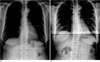 | Fig. 1Measuring Cobb's angle by Cobb method of 44 years old female patient.
A. Preoperative chest plane radiograph.
B. Whole spine standing anteroposterior view after 4 years.
|
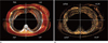 | Fig. 2Measured volume of the chest wall by 3D reconstructed volume rendering image of chest MDCT.
A. Whole chest wall volume is divided into 4 segments by right anterior (RtA), left anterior (LtA), right posterior (RtP), left posterior (LtP).
B. The muscle volume of chest wall is measured with 20-80 HU threshold (mRtA, mLtA, mRtP, mLtP).
Note.-MDCT = multi-detector computed tomography
|

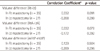
Table 2
Chest Wall Volume Differences in Mastectomy Patients by Chest MDCT with 3D Volume Reconstruction
![]()

References
1. Jemal A, Siegel R, Ward E, Hao Y, Xu J, Murray T, et al. Cancer Statistics, 2008. CA Cancer J Clin. 2008; 58:71–96.
2. Ministry for Health, Welfare and Family Affairs. Annual Report of cancer incidence (2007), cancer prevalence (2007) and survival (1993-2007) in Korea. Seoul: Ministry for Health, Welfare and Family Affairs;2009.
3. Won YJ, Sung J, Jung KW, Kong HJ, Park S, Shin HR, et al. Nationwide cancer incidence in Korea, 2003-2005. Cancer Res Treat. 2009; 41:122–131.
4. Kuehn T, Klauss W, Darsow M, Regele S, Flock F, Maiterth C, et al. Long-term morbidity following axillary dissection in breast cancer patient--clinical assessment, significance for life quality and the impact of demographic, oncologic and therapeutic factors. Breast Cancer Res Treat. 2000; 64:275–286.
5. Bower JE, Ganz PA, Desmond KA, Bernaards C, Rowland JH, Meyerowitz BE, et al. Fatigue in long-term breast carcinoma survivors: a longitudinal investigation. Cancer. 2006; 106:751–758.
6. Bendz I, Fagevik Olsén M. Evaluation of immediate versus delayed shoulder exercise after breast cancer surgery including lymph node dissection--a randomised controlled trial. Breast. 2002; 11:241–248.
7. Wingate L. Efficacy of physical therapy for patients who have undergone mastectomies. A prospective study. Phys Ther. 1985; 65:896–900.
8. Aebi M. The adult scoliosis. Eur Spine J. 2005; 14:925–948.
9. Lee SW, Lee TJ, Lee SW. Does unilateral mastectomy cause scoliosis? J Korean Soc Plast Reconstr Surg. 2008; 35:279–282.
10. Shamley D, Srinaganathan R, Oskrochi R, Lascurain-Aguirrebeña I, Sugden E. Three-dimensional scapulothoracic motion following treatment for breast cancer. Breast Cancer Res Treat. 2009; 118:315–322.
11. Cheville AL, Tchou J. Barriers to rehabilitation following surgery for primary breast cancer. J Surg Oncol. 2007; 95:409–418.
12. Hagay C, Cherel PJ, de Maulmont CE, Plantet MM, Gilles R, Floiras JL, et al. Contrast-enhanced CT: value for diagnosing local breast cancer recurrence after conservative treatment. Radiology. 1996; 200:631–638.
13. Lindfors KK, Meyer JE, Busse PM, Kopans DB, Munzenrider JE, Sawicka JM. CT evaluation of local and regional breast cancer recurrence. AJR Am J Roentgenol. 1985; 145:833–837.
 XML Download