

 PDF
PDF ePub
ePub Citation
 Print
Print


Abstract
Purpose
We evaluated the utility of iopamidol-based nonionic contrast media (Pamiray®370) for cardiac CT, with assessment of its image quality and safety.
Materials and Methods
The study included 100 patients who underwent cardiac CT with Pamiray®370 (experimental group), and 100 patientswho underwent cardiac CT with Ultravist®370 (control group). A comparison of the image qualities and degree of vascular contrast enhancement was made between the two groups and evaluated statistically by an independent t-test. Changes in vital signs and adverse events during cardiac CT were evaluated in the experimental group.
Results
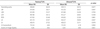
There were no statistically significant differences in the image quality (image quality score in experimental group vs. control group: 4.26 ± 0.63 vs. 4.24 ± 0.62), and mean attenuation values at the coronary arteries(p > 0.05) between two groups. For the experimental group, 12% experienced adverse events, including mild and transient reactions such as dizziness (7%), nausea (4%), and fatigue (1%). Further, 94% of patients complained of mild to moderate febrile sense just after contrast agent administration, which spontaneously disappeared within 3 minutes without any specific management.
Figures and Tables
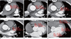
Fig. 1
Assessment of the attenuation value (HU) at the ascending aorta, coronary vessels, and left cardiac chamber. Attenuation values were assessed at the regions of interest (ROI), which were positioned at the center of the ascending aorta (A), at the orifice levels of the left main coronary artery (B), left anterior descending artery (C), left circumflex artery (D) and right coronary artery (E), at the mid-portions of left atrium, left ventricle, and left ventricular myocardium (F).

References
1. Katayama H, Yamaguchi K, Kozuka T, Takashima T, Seez P, Matsuura K. Adverse reactions to ionic and nonionic contrast media. A report from the Japanese Committee on the Safety of Contrast Media. Radiology. 1990; 175:621–628.
2. Korn WT, Bettmann MA. Low-osmolality versus high-osmolality contrast material. Curr Opin Radiol. 1992; 4:9–15.
3. Bettmann MA, Heeren T, Greenfield A, Goudey C. Adverse events with radiographic contrast agents: results of the SCVIR Contrast Agent Registry. Radiology. 1997; 203:611–620.
4. Grainger RG. Osmolality of intravascular radiological contrast media. Br J Radiol. 1980; 53:739–746.
5. Grainger RG. Intravascular contrast media. Br J Radiol. 1982; 55:544.
6. McClennan BL, Stolberg HO. Intravascular contrast media. Ionic versus nonionic: current status. Radiol Clin North Am. 1991; 29:437–454.
7. Stolberg HO, McClennan BL. Ionic versus nonionic contrast use. Curr Probl Diagn Radiol. 1991; 20:47–88.
8. Wolf GL, Arenson RL, Cross AP. A prospective trial of ionic vs nonionic contrast agents in routine clinical practice: comparison of adverse effects. AJR Am J Roentgenol. 1989; 152:939–944.
9. Amiel M, Moll T, Revel D, Corot C, Touboul T, Kirkorian G, et al. Comparison of the electrophysiologic effects of ioxaglate and iopamidol during selective coronary arteriography. Invest Radiol. 1990; 25:Suppl 1. S141–S143.
10. Bannon KR, Braun IF, Pinto RS, Manuell M, Sudilovsky A, Kricheff II. Comparison of radiographic quality and adverse reactions in myelography with iopamidol and metrizamide. AJNR Am J Neuroradiol. 1983; 4:312–313.
11. Conroy RM, Bjartveit K, Sheppick A, Long U, Masterson J. Iodixanol in intravenous urography: a comparison of iodixanol 270 mgI/ml, iodixanol 320 mgI/ml and iopamidol 300 mgI/ml (NIOPAM). Clin Radiol. 1994; 49:337–340.
12. Heuschmid M, Küttner A, Flohr T, Wildberger JE, Lell M, Kopp AF, et al. [Visualization of coronary arteries in CT as assessed by a new 16 slice technology and reduced gantry rotation time: first experiences]. Rofo. 2002; 174:721–724.
13. Cademartiri F, van der Lugt A, Luccichenti G, Pavone P, Krestin GP. Parameters affecting bolus geometry in CTA: a review. J Comput Assist Tomogr. 2002; 26:598–607.
14. Bae KT, Heiken JP, Brink JA. Aortic and hepatic peak enhancement at CT: effect of contrast medium injection rate--pharmacokinetic analysis and experimental porcine model. Radiology. 1998; 206:455–464.
15. Fleischmann D, Rubin GD, Bankier AA, Hittmair K. Improved uniformity of aortic enhancement with customized contrast medium injection protocols at CT angiography. Radiology. 2000; 214:363–371.
16. Lee Y, Lee J, Lee HJ, Park J. The efficacy of Iopamidol (Pamiray®370) in aortic and extremity CT angiography. J Korean Soc Radiol. 2010; 62:23–28.
17. Cademartiri F, Mollet NR, van der, McFadden EP, Stijnen T, de Feyter PJ, et al. Intravenous contrast material administration at helical 16-detector row CT coronary angiography: effect of iodine concentration on vascular attenuation. Radiology. 2005; 236:661–665.
18. Park SH, Suh SH, Kim J, Kim EY, Kim DJ, Lee SK, et al. Cinical application of Iopamidol (Pamiray®300) for cerebral angiography. J Korean Radiol Soc. 2007; 57:121–127.
19. Dawson P. Chemotoxicity of contrast media and clinical adverse effects: a review. Invest Radiol. 1985; 20:S84–S91.
20. Manke C, Marcus C, Page A, Puey J, Batakis O, Fog A. Pain in femoral arteriography. A double-blind, randomized, clinical study comparing safety and efficacy of the iso-osmolar iodixanol 270 mgI/ml and the low-osmolar iomeprol 300 mgI/ml in 9 European centers. Acta Radiol. 2003; 44:590–596.
21. Pugh ND, Sissons GR, Ruttley MS, Berg KJ, Nossen JO, Eide H. Iodixanol in femoral arteriography (phase III): a comparative double-blind parallel trial between iodixanol and iopromide. Clin Radiol. 1993; 47:96–99.
22. Faykus MH Jr, Cope C, Athanasoulis C, Druy EM, Hedgcock M, Miller FJ, et al. Double-blind study of the safety, tolerance, and diagnostic efficacy of iopromide as compared with iopamidol and iohexol in patients requiring aortography and visceral angiography. Invest Radiol. 1994; 29:Suppl 1. S98–S101. discussion S106.
23. Mortelé KJ, Oliva MR, Ondategui S, Ros PR, Silverman SG. Universal use of nonionic iodinated contrast medium for CT: evaluation of safety in a large urban teaching hospital. AJR Am J Roentgenol. 2005; 184:31–34.
 XML Download