

 PDF
PDF ePub
ePub Citation
 Print
Print


INTRODUCTION
The incidence of thyroid nodules in children and adolescents is estimated to be between 1% and 2% (1). However, because of the detection of incidental and subclinical thyroid nodules using diagnostic imaging modalities, this incidence may be increasing. The risk of malignancy of thyroid nodules in children ranges between 14 to 40%, which is much higher than that of adults (2). The estimated annual incidence of primary thyroid carcinoma in this population worldwide is 0.5-10 cases per million (2). In Korea, thyroid cancer is the 11th most commonly diagnosed cancer in children under fourteen years old and the 10th most common cancer in girls (http://www.ncc.re.kr) (3).
In children, thyroid cancer tends to present at a more advanced stage than in adults, with a higher frequency of lymph node and pulmonary metastases (4). Distant metastasis is much less common than regional lymph node involvement, with the lung being the most common site of distant metastasis (56).
Several reports have suggested that papillary thyroid carcinomas (PTCs) follow a more favorable course in young patients (< 21 years of age) (7). The reasons for this variation between children and adults are unknown, but are generally thought to be due to differences in mutations and oncogene expression in the thyroid (8). PTC is frequently associated with genetic alterations leading to the activation of the mitogen-activated protein kinase signaling pathway (910). A BRAFV600E mutation at nucleotide position 1799 is a transversion of a thymine to an adenine, resulting in the conversion of a valine to a glutamate. The BRAFV600E mutation is intrinsically associated with increased progression and aggressiveness of PTC. The reported frequency of a specific BRAFV600E mutation in a PTC series ranged from 36% to 65% (11). However, the prevalence of the BRAFV600E mutation in PTC in Korea is much higher (70-83%) than that of Western countries (12). Moreover, compared to rearranged during transfection/Papillary Thyroid Carcinomas, the BRAFV600E abnormality is a more common genetic alteration, occurring in half of sporadic PTCs (13).
Ultrasonography (US) is commonly used for the evaluation of thyroid nodules. However, US and CT reports regarding the identification and features of suspicious nodules in children have been limited (1415).
The purpose of the present study was to review the clinical and imaging features of pediatric thyroid cancer and the status of molecular markers for the BRAFV600E mutation in PTC, as well as analyzing the US and CT scan features of histopathologically confirmed thyroid cancer.
MATERIALS AND METHODS
This study was approved by the institutional review board with a waiver of informed consent.
Patients
Thirteen patients under eighteen years of age with histopathologically confirmed thyroid cancer from April 2003 to November 2009 were enrolled in the present study.
Medical records and imaging features were reviewed. The data collected included gender, age at diagnosis, presenting symptoms, lymph node involvement, distant metastases, tumor node metastasis (TNM)/pathological TNM classification, type of surgery performed, histological type, and molecular marker for the BRAFV600E mutation associated with PTC. The TNM staging adopted in the present study was the American Joint Committee on Cancer (AJCC) classification for thyroid cancer (2010) (16).
Imaging Techniques and Image Interpretation Ultrasonography, FNA, and BRAFV600E Analysis
Nine of 13 patients underwent US examination using an IU22 US, HDI 5,000 with a 12 MHz linear array transducer (Philips Medical Systems, Bothell, WA, USA), and a LOGIQ 700 MR system with a 10 MHz linear array transducer (GE Healthcare, Milwaukee, WI, USA). US findings were analyzed for location, presence of a nodule, echotexture, echogenicity, calcifications, margin, shape, intranodular vascularity on color Doppler images and abnormal lymph nodes in the lateral neck. US findings were analyzed based on the "Guidelines for the Thyroid US" by the Thyroid Study Group, Korean Society of Neuro- and Head and Neck Radiology (17). Nodule echogenicity was assessed according to the surrounding thyroid tissue and was classified as hyperechoic, isoechoic, hypoechoic, or markedly hypoechoic. The nodule echotexture was classified as homogeneous or heterogeneous. The margin of the nodule was classified as smooth or irregular. The shape of the nodule was categorized as either ovoid to round or taller than wide. The presence of calcifications and abnormal lymph nodes in the lateral neck was also recorded. Calcifications < 1 mm were classified as microcalcifications, and those > 1 mm were classified as macrocalcifications. Abnormal lymph node criteria included enlarged size (> 1.5 cm in the longest diameter at neck levels I and II, > 1.0 cm in the longest diameter at other neck node levels), spherical in shape (longitudinal/transverse diameter is < 1.5 cm), central necrosis, calcification, and chaotic vessels on a color Doppler image.
During color Doppler US examination, the presence of intra nodular blood flow was evaluated for all lesions. A thyroid gland was considered to be goiterous when its volume exceeded the 97th percentile of the International Council for Control of Iodine Deficiency Disorders reference value (18).
BRAFV600E mutations were analyzed in ten patients. A 22- gauge needle attached to a 10-mL disposable plastic syringe was used to perform ultrasound-guided fine needle aspiration biopsy. Each lesion was aspirated at least twice and the specimen for analysis of the BRAFV600E mutation was obtained separately. The specimen was rinsed with 1 mL of normal saline, and then the wash-out was subjected to BRAFV600E mutation analysis in three patients; another eight patients were evaluated for BRAFV600E with samples from paraffin embedded blocks. Dual-priming oligonucleotide-based multipex polymerase chain reaction (PCR) amplification methods were used for the identification of mutations from a portion of the BRAFV600E gene using Seeplex BRAF ACE Detection (Seegene, Inc., Seoul, Korea). Paraffin-embedded thyroid cancer tissue samples were examined twice, including DNA isolation and PCR amplification for the BRAFV600E mutation (WH Lee).
Contrast-Enhanced Neck CT
Twelve of 13 patients underwent CT examination. A multi-detector row CT (MDCT) unit (Somatom Sensation 16; Siemens Medical Solutions, Erlangen, Germany) with 16 detector rows (n = 4); a spiral CT unit (n = 6) (HiSpeed Advantage; GE Healthcare, Milwaukee, WI, USA), and an MDCT unit with 64 detector rows (n = 1) (LightSpeed VCT; GE Healthcare, Milwaukee, WI, USA) were used. One patient underwent CT at an outside hospital. Typical contrast-enhanced CT imaging protocols for thyroid disease included unenhanced CT and contrast-enhanced CT images at 80 kVp and 60 mAs; field of view = 23 cm; collimation = 0.625 to 1.25 mm; and pitch = 0.9 to 1.3. Adaptation of the tube current corresponding to the body weight is used as a dose-saving CT protocol. For contrast-enhanced CT scans, 1.2-1.5 mL/kg of an iodinated contrast agent (Ultravist 360; Schering, Berlin, Germany), ranging from 0.3 to 3 mL/s, was injected, CT characteristics of the lesions, including attenuation, compared with the surrounding normal thyroid gland on pre- and post-contrast enhancement imaging, presence of calcifications, and measured degree of enhancement, were assessed. The degree of enhancement was calculated in Hounsfield units using the following formula: [(Density post-Density pre)/Density pre] × 100%. The region of interest is positioned centrally with respect to the nodule or the goiterous enlarged lesions.
Eleven out of 13 patients underwent surgery; two patients were lost to follow up. All neoplastic lesions were staged according to the AJCC classification for thyroid cancer (2010) (16).
RESULTS
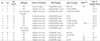
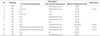
The series of patients consisted of ten girls and three boys, ranging in age 6 to 18 years old (mean, 15.5 years old). The clinical information is summarized in Table 1. No patient had a history of radiation exposure. Eleven patients presented with a palpable neck mass, one patient presented with vocal cord palsy, and one patient presented with an incidentally detected thyroid mass on a chest CT scan, which was performed as a result of chest pain. The pathological diagnosis was confirmed at surgery (n = 11) or during fine needle aspiration biopsy (n = 2). Two patients who underwent hemithyroidectomy did not undergo preoperative US; one had nodular hyperplasia with intrathyroidal PTC, and the other had minimally invasive follicular thyroid cancer (FTC). Seven patients underwent a neck dissection due to metastatic lymphadenopathy. In total, eleven patients (84.6%) were diagnosed with PTC, of which all cases were of the classic type, and two patients (15.4%) were diagnosed with minimally invasive FTC. Metastasis was detected in nine patients; lymph node involvement was observed in eight (61.5%), and lung metastasis was detected in one (7.7%) BRAFV600E mutations were detected in three (30.0%) of 10 PTC patients.
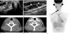
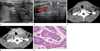
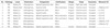
The US features of nine patients are summarized in Table 2. Seven patients had nodular lesions (Fig. 1) and the remaining two patients showed goiterous enlargement of the thyroid glands with microcalcifications (Fig. 2). All seven nodular lesions were solid and heterogeneous in echotexture. All six PTC nodules showed malignant US findings according to the "Guidelines for the Thyroid US" of the Thyroid Study Group, Korean Society of Neuro- and Head and Neck Radiology (17) [four findings (n = 2); three findings (n = 1); two findings (n = 3)]. One minimally invasive FTC demonstrated indeterminate nodule on US. The average nodule size had a maximum diameter of 1.93 cm (diameter range; 1.5-2.7 cm). Echogenicity of nodules was marked hypoechoic (3/7; 42.9%) (Fig. 1A), hypoechoic (1/7; 14.3%), isoechoic (2/7; 28.6%), or hyperechoic (1/7; 14.3%). Calcifications (micro- or macro-) were found in eight (88.9%) patients; the margin was irregular in six (85.7%) patients, all of which had PTC, and smooth in one patient with FTC. A taller than wide shape was found in two (28.6%) patients with PTC (Fig. 1A). All lesions revealed increased vascularity within the nodules or a goiterous enlarged gland on a color Doppler image. In two patients having diffuse enlargement with calcifications, the tumor replaced the entire gland. Abnormal neck lymph nodes were found in seven (77.8%) patients (Fig. 1B).
Twelve patients underwent CT examination. Among ten nodular lesions found, eight were low attenuation compared with the surrounding normal thyroid gland, and one had microcalcifications. The other two patients with underlying lymphocytic thyroiditis showed diffuse low attenuation on the pre-contrast scan, which was similar to that of the adjacent strap muscle and the nodular lesions were iso to low attenuation. The two remaining patients showed a goiterous enlarged gland with microcalcifications (Fig. 2). Eleven nodular lesions showed low attenuation compared with well enhancing surrounding thyroid glands, which were detected more easily on post-contrast enhanced scans than on pre-contrast scans (Fig. 1C, D). The degree of enhancement ranged from 80% to 406% (mean: 166.9%) in 11 patients (one patient underwent a CT scan at an outside hospital; thus, the degree of enhancement could not be calculated). One nodule from the follicular thyroid cancer in our study was well enhanced (406%); the CT features are summarized in Table 3.
DISCUSSION
Gender and age distribution in the present study were similar to those of other studies. Additionally, nine patients showed high T-staging; T3 (n = 5; 38.5%) and T4 (n = 4; 30.8%), and metastatic lymphadenopathy in the lateral neck (n = 8; 61.5%). These results are consistent with those of previous reports showing that pediatric thyroid cancer tends to present at a more advanced stage than that of adults (4). In a long term follow-up study reported by Zimmerman et al. (5), the incidence of distant metastasis was estimated to be 6.9% in children, with lymph node involvement in 89.7%.
PTC is the most common thyroid cancer in children; the prognosis is more favorable in young patients (7). In our study, the BRAFV600E mutation was detected in three of ten patients (30.0%) with PTC which was a much lower frequency than what was found in Korean adults (70-83%) (12); however, it is higher than that of other reports on pediatric thyroid cancer (9). The BRAFV600E mutation is intrinsically associated with increased progression and aggressiveness of PTC. Similarly, the BRAFV600E mutation has a similarly high predictive value for PTC recurrence (1113). Pediatric PTC is highly curable and rarely recurs if treated with appropriate surgery with adjunctive radioiodine ablation, even if the initial disease is extensive and associated with extrathyroidal invasion, lymph node metastasis, or lung metastasis (1920). This is consistent with the fact that the BRAFV600E mutation rarely occurrs in pediatric PTC (910). In this small study population, we cannot explain the relevance of imaging findings in the prediction of prognosis. The low rate of the BRAFV600E mutation in our study supports the concept that pediatric thyroid cancer is almost always differentiated and sensitive to radioiodine treatment, leading to a better prognosis.
Using gray-scale and power Doppler imaging, Lyshchik et al. (15) reported in their study of US features of thyroid cancer in children that only included focal nodular lesions of the thyroid. Although our study population was small, two out of nine patients (22.2%) showed US findings of diffuse enlargement with microcalcifications, which were confirmed to be classical PTC. The thyroid gland was replaced by tumor cells in these cases. Nodules were observed in patients who had underlying lymphocytic thyroiditis; one patient showed a typical malignant nodule, and the other, who presented with hyperthyroidism (thyroid uptake 7.2%; normal range 2-4%), showed an oval hyperechoic nodule with numerous microcalcifications. Thus, patients with a diffusely enlarged thyroid with microcalcifications should undergo FNA and BRAFV600E analysis. Regarding nodules, US findings of nodules did not differ from malignant nodules in adults. All PTC nodules were suspicious malignant nodules according to the "Guidelines for the Thyroid US" by the Thyroid Study Group, Korean Society of Neuro- and Head and Neck Radiology. In the present study, calcifications (88.9%, 8/9) and intranodular vascularity (100%) on color Doppler images were frequent findings on US, whereas 33.3% (4/12) of the patients showed calcifications on CT scan. This difference was probably due to that fact that microcalcifications are more easily detected on US.
Because of the negative effect of postoperative I-131 ablation, the preoperative use of iodinated contrast agents in patients with thyroid cancer is discouraged (21). However, in the present study, contrast enhanced CT scans did not alter or affect patient management, because patients were required to wait until a therapeutic room for iodine ablation therapy became available. US can evaluate thyroid abnormalities and lymphadenopathy. CT scans are useful for the evaluation of lymph nodes at the level of VI or retropharyngeal nodes, which are limited areas on US. Furthermore, despite a very high accuracy of US according to a per patient analysis, the sensitivity of CT according to per level analysis is superior to that of US, so CT may play a complementary role in the determination of the surgical extent in selected patients with thyroid cancer (22). In our study, contrast-enhanced CT scans showed low attenuation nodular lesions in 11 patients (11/12) in comparison to well-enhanced surrounding thyroid glands. In one patient who underwent CT in an outside hospital, the degree of enhancement could not be calculated; the degree of enhancement ranged from 80 to 406% (mean, 166.9%) in 11 patients. Because this study included a small number of patients, a subsequent study comparing benign nodules in a larger group of patients will be required.
In summary, pediatric thyroid cancer tends to present with high T-stage. BRAFV600E mutations were detected in 30.0% of cases, which is a lower frequency than the frequency found in adults. US features showed a goiterous enlargement with calcification (n = 2; 22.2%) or a nodular lesion (n = 7; 77.8%). We suggest that a diffusely enlarged thyroid with microcalcifications should be evaluated with FNA. On contrast-enhanced CT, a low attenuation nodule was the main finding.
 XML Download