

 PDF
PDF ePub
ePub Citation
 Print
Print


Mediastinitis refers to inflammation of the mediastinal structures that lie between the right and left pleura between the thoracic inlet and the diaphragm. Mediastinitis can be divided into the acute and chronic forms according to the etiology and the clinical course. Acute suppurative mediastinitis is an uncommon and life threatening condition (1).
The main causes of acute mediastinitis include esophageal perforation, post-operative complications and the spread of an adjacent infection. Since the development of antibiotics, acute mediastinitis not caused by trauma or surgery is rare (2).
Therefore, we report here on a case of a ten-year-old female patient with an acute mediastinal abscess that occurred in the absence of trauma, surgery or deep neck infection.
Case Report
A 10-year-old girl presented with fever, sore throat and odynophagia that she'd had for five days. She had no previous history of surgery or medical illness, including allergies and tuberculosis, and she had no history of swallowing fish bones.
Upon examination, the patient's temperature was 39.7 ℃ and her heart rate was 144 beats/min. The laboratory data revealed a white blood cell count of 12,450 mL-1 (normal range: 4,000-10,800 mL-1) with 86% neutrophils (normal range: 40-73%), an ESR of 88 mm/Hr (normal range: 0-20 mm/Hr) and a CRP level of 370.8 mg/L (normal range: 0.1-6.0 mg/L).
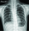
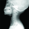
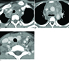
Chest radiograph showed superior mediastinal widening (Fig. 1), but the lateral neck view was unremarkable with normal retropharyngeal and retrotracheal soft tissue thicknesses (Fig. 2). On the non-enhanced computed tomography (CT), we found no calcification or free air, although the contrast enhanced CT images revealed extensive inflammation and abscesses involving the entire mediastinum (Figs. 3A, B). The inflammatory lesions reached to the level of the left thyroid bed (Fig. 3C), but there was no evidence of a deep neck infection. There were no abnormalities in the bony structures or lung parenchyma. The trachea and esophagus were surrounded by abscesses and inflammation, but esophageal wall thickening, an extraluminal air collection and pneumohydrothorax were not observed.
Sternotomy with incision and drainage was performed. A large amount of greenish pus was drained, and mediastinitis with abscess formation and a massive infiltration of acute and chronic non-specific inflammatory cells were pathologically confirmed. A bacteriologic study showed gram positive cocci.
Discussion
Mediastinitis is defined as inflammation in the mediastinal structures (1). Although mediastinitis is a relatively uncommon disease, it is a life-threatening condition with a mortality rate of up to 40% (23). Acute suppurative mediastinits is usually caused by chest surgery or esophageal perforation. Additionally, tracheal perforation, endoscopic trauma, foreign body ingestion, carcinomas, mediastinal lymph node infection, an extended infection from adjacent structures such as clavicular osteomyelitis and descending spread of cervical infection can also induce mediastinitis (14).
Chills, fever, tachycardia, pain and sepsis are the common clinical symptoms of the disease. Making a fast and accurate diagnosis and administering appropriate treatment are critical. The patient's history, clinical symptoms and physical findings, and plain radiography and CT are used for making the proper diagnosis. Widening of the mediastinum, associated pleural disease, air-bubble formation inside the mediastinum and a mediastinal soft tissue mass or an air-fluid level are common plain radiographic findings (5). However, these findings are also non-specific, and it is difficult to recognize the exact extent of the disease using only plain radiographs. Therefore, CT is the method of choice for diagnosing acute mediastinitis. CT is useful not only for making the diagnosis, but also for the exact evaluation of the location and extent of the disease. It also helps in identifying any coexisting diseases (6).
The most common CT findings of acute mediastintis are loculated mediastinal fluid collections, air bubbles, increased attenuation of the mediastinal fat, pleural and pericardial effusion and enlarged mediastinal lymph nodes (147). The CT findings may be variable depending on the underlying causes. Acute mediastinitis caused by esophageal perforation is accompanied with esophageal wall thickening, an extraluminal air collection and pneumohydrothorax, while mediastinitis caused by cardiac or mediastinal operations is usually accompanied by sternal abnormalities (57). Descending necrotizing mediastinitis (DNM) is an unusual mediastinitis that spreads from a head or neck infection. Widening of the superior mediastinum, increased density of the adipose tissue, myositis, cervical lymphadenopathy, a mediastinal fluid collection, a pleural/pericardial fluid collection and vascular thrombosis are the major CT features of DNM (189).
In our case, the patient had no history of surgery or trauma. On the CT scan, severe mediastinitis with abscesses was found only below the thyroid bed, and there was no evidence of infection involving the head or neck. To the best of our knowledge, no cases of spontaneous mediastinal abscess in children have been reported in the English medical literature.
Acute mediastinitis caused by polymicrobial infection requires immediate and prompt broad-spectrum antibiotic therapy and surgical treatment for obtaining a better prognosis. As in our case, early surgical mediastinotomy is preferred, but minimally-invasive thoracic drainage could be performed with good results in the cases with an early diagnosis (10).
 XML Download