

 PDF
PDF ePub
ePub Citation
 Print
Print


Collagenous fibroma is a recently defined entity that was first described by Evans in 1995 as desmoplatic fibroblastoma and this tumor was renamed 1 year later by Nielsen et al. (12). Since its initial description in 1995, fewer than 100 cases have been reported with the largest series having been published by Miettinen and Fetsch (3). Collagenous fibroma is a benign fibrous soft tissue tumor that typically arises in the subcutaneous tissue or skeletal muscle of adults. This tumor typically presents as a slowly growing, painless mass. This tumor is found in a people of all ages, but it is most common in the fifth and sixth decades of life. Men are affected four times more commonly than women. This entity has been reported in various locations, including the upper extremities, lower extremities, posterior neck, upper back, abdominal wall, hip joint and head. Only a few case reports have described the MRI features of this tumor. We herein report on a case of a collagenous fibroma in the finger and we place emphasis on the MRI findings with the pathological correlation.
Case Report
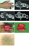
A 54-year-old man was admitted to our hospital with a slowly growing, painless mass in the dorsoradial aspect of the proximal phalanx of the right 3rd finger. He had a history of fracture of the distal phalanx of the 3rd finger 5 years ago. Two year later, he had felt the slowly growing mass. Physical examination showed an approximately 4 × 6 cm2 firm, fixed mass without tenderness (Fig. 1A).
An MRI study was done. On the T1-weighted images, the mass was well-circumscribed, and it had iso-signal intensity compared with muscle (Fig. 1B). On the T2-weighted images, the mass had diffuse low signal intensity with focal high signal intensity (Fig. 1C). The contrast-enhanced T1-weighted fat-saturated images showed mild heterogeneous enhancement with focal irregular areas of non-enhancement (Fig. 1D).
The tumor was removed by surgical excision. The tumor was located in the subcutaneous tissue of the 3rd finger. The tumor was loosely attached to the tendon sheath, but it had not invaded the extensor and flexor tendons (Fig. 1E). Macroscopically, the tumor appeared as a multilobulated, firm, whitish mass without gross necrosis, and it measured 5.5 × 5.0 × 2.5 cm3 (Fig. 1F). Microscopically, the lesion was hypocellular and composed of fibroblast/myofibroblast cells dispersed in a collagenous-rich stroma (Fig. 1G). Cytological atypia, mitotic figures, areas of necrosis, calcifications and inflammatory cells were not seen. Immunohistochemical staining showed the tumor cells to be focally and weakly positive for smooth muscle actin and they were negative for S100, CD 34 and CD 68. Thus, fibroma of the tendon sheath and nerve sheath tumor were excluded from the differential diagnosis.
There has been no local tumor recurrence at the 6 month postoperative follow-up examination.
Discussion
Collagenous fibroma characteristically presents as slowly growing, non-tender masses that have developed over a long duration. The tumor has a male predominance and a peak incidence in the fifth and sixth decades of life. This tumor usually occurs in the subcutaneous tissue, but approximately 25% of the tumors involve skeletal muscle. The tumor has a wide anatomic distribution, but it mainly affects the extremities. The tumor typically infiltrates fat and skeletal muscle, and this has been observed in up to 51% of cases (3). The tumors range in size from 1 to 20 cm. Histologically, the tumor is hypocellular and it consists of stellate and spindle-shaped fibroblastic cells that are widely separated by a collagenous to fibromyxoid matrix (134).
Treatment of collagenous fibroma is surgical excision and there have been no reported incidences of local recurrence or metastases (35).
Evans postulated that collagenous fibromas might be either neoplasm or a reactive condition (1). A history of antecedent trauma has been noted in only three cases of collagenous fibroma (35). Our report reinforces the possibility that fibrous proliferation subsequent to trauma may contribute to the development of these tumors.
There have been seven previous case reports regarding the MRI features of collagenous fibroma. Among these cases, four case reports had the contrast-enhanced T1-weighted images (5678). On MRI, collagenous fibroma has a low signal on both the T1-weighted and T2-weighted images. The low signal intensity of the mass on both the T1-weighted and T2-weighted images is attributed to the low cellularity of the mass in a background of abundant collagen. The areas of persistently low signal intensity on the contrast-enhanced T1-weighted images correspond to regions of dense, relatively acellular collagen matrix (67). Our case was consistent with the previous case reports.
Most soft-tissue masses have high signal intensity on T2-weighted images. The soft-tissue masses with low SI on the T2-weighted images include fibroma of the tendon sheath, neurofibroma, cicatrical fibroma, malignant fibrous histiocytoma, aggressive fibromatosis and calcifying fibrous pseudotumor (59). Collagenous fibroma may be misdiagnosed as one of these soft tissue tumors.
In summary, we report here on a collagenous fibroma of the finger that presented with a history of antecedent trauma to the finger. Collagenous fibroma should be included in the differential diagnosis of a well-circumscribed lesion with low signal intensity seen on both the T1-weighted and T2-weighted images and the lesion shows minimal enhancement.
 XML Download