

 PDF
PDF ePub
ePub Citation
 Print
Print


Abstract
Purpose
To evaluate the surgical outcomes of fibroepithelial lesion with cellular stroma (FELCS) diagnosed at sonography guided core needle biopsy of breast masses, and to determine whether the clinical and imaging features of this lesion could predict the presence of a phyllodes tumor.
Materials and Methods
We retrospectively reviewed the pathologic results of sonography guided core needle biopsy of solid breast masses. A total of 55 FELCS diagnosed with this procedure that underwent subsequent surgical excision were included in this study; their medical records and radiologic images were retrospectively reviewed.
Results
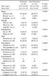
The results of the surgical excision revealed 22 (40%) phyllodes tumors and 33 (60%) non-phyllodes tumors: 30 (54.6%) fibroadenomas, 1 (1.8%) adenosis, 1 (1.8%) fibrocystic changes and 1 (1.8%) fibroadenomatous hyperplasia. Lesion size and patient age were significantly different between phyllodes tumors and non-phyllodes tumors groups (32.2 ± 14.07 mm/22.4 ± 13.64 mm, p=0.0078, 43.5 ± 11.60 years/36.5 ± 10.25 years, p=0.0207). Among the sonographic features, only cleft was significantly more visible in phyllodes tumors than in non-phyllodes tumors (n=14 (70%)/n=6 (30%), p=0.0016).
Figures and Tables
Fig. 3
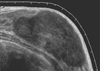
18-year-old woman with a palpable mass diagnosed as fibroepithelial lesion with cellular stroma at ultrasonography guided core biopsy.
A. Ultrasonography shows a 41 mm sized, irregular, not-circumscribed and hypoechoic mass with clefts categorized as Category 4. It was confirmed as benign phyllodes tumor at surgical excision.
B. Photomicrograph of core needle biopsy specimen shows increased stromal cellularity (arrows) and normal ducts (arrowheads) (H and E, ×40).
C. Photomicrograph of surgical excision specimen shows epithelial lining stroma with significantly increased cellularity, which is more compatible with phyllodes tumor than fibroadenoma (H and E, ×40).
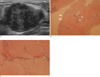
Fig. 4
32-year-old woman with a palpable mass diagnosed as fibroepithelial lesion with cellular stroma at ultrasonography guided core biopsy ultrasonogram shows a 34 mm sized, oval, not-circumscribed and hypoechoic mass with clefts was categorized as Category 4. It was confirmed as phyllodes tumor at surgical excision.
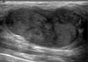
Fig. 5
19-year-old woman with a palpable mass diagnosed as fibroepithelial lesion with cellular stroma at ultrasonography guided core biopsy Ultrasonogram shows a 60 mm sized, oval, circumscribed and hypoechoic mass categorized as Category 3. It was confirmed as fibroadenoma at surgical excision.

Acknowledgement
This research was supported by the Basic Science Research Program through the National Research Foundation of Korea (NRF) funded by the Ministry of Education, Science and Technology (2009-0067048).
References
1. Jacobs TW, Chen YY, Guinee DG, Holden JA, Cha I, Bauermeister DE, et al. Fibroepithelial lesions with cellular stroma on breast core needle biopsy: are there predictors of outcome on surgical excision? . 2005; 124:342–354.
2. Rosen PP. Fibroepithelial lesions. Rosen's breast pathology. 2nd ed. Philadelphia: Lipincott Williams & Williams;2001. p. 163–200.
3. Shousha S. Issues in the interpretation of breast core biopsies. Int J Surg Pathol. 2003; 11:167–176.
4. Bode MK, Rissanen T, Apaja-Sarkkinen M. Ultrasonography and core needle biopsy in the differential diagnosis of fibroadenoma and tumor phyllodes. Acta Radiol. 2007; 48:708–713.
5. Jacobs TW, Connolly JL, Schnitt SJ. Nonmalignant lesions in breast core needle biopsies to excise or not to excise? Am J Surg Pathol. 2002; 26:1095–1110.
6. Dershaw DD, Morris Ea, Liberman L, Abramson AF. Nondiagnostic stereotaxic core breast biopsy: results of rebiopsy. Radiology. 1996; 198:323–325.
7. Meyer JE, Smith DN, Lester SC, DiPiro PJ, Denison CM, Harvey SC, et al. Large-needle core biopsy: nonmalignant breast abnormalities evaluated with surgical excision or repeat core biopsy. Radiology. 1998; 206:717–720.
8. Komenaka IK, EL-Tammer M, Pile-Spellman E, Hibshoosh H. Core needle biopsy as a diagnostic tool to differentiate phyllodes tumor from fibroadenoma. Arch Surg. 2003; 138:987–990.
9. Jung HK, Kim EK, Go KH, Rho JY. Phyllodes tumors or fibroepithelial lesions with cellular stroma of breast diagnosed at sonographically guided core needle biopsy: comparison between results on excision biopsy and sonographic findings. J Korean Soc Ultrasound Med. 2011; 30:45–53.
10. Stravros AT. Atypical, high-risk, premalignant, and locally aggressive lesions. In : Stavros AT, editor. Breast Utrasound. Philadelphia: Lippincott, Williams & Wilkins;2001. p. 695–701.
11. Reinfuss M, Mitus J, Duda K, Stelmach A, Rys J, Smolak K. The treatment and prognosis of patients with phyllodes tumor of the breast: an analysis of 170 cases. Cancer. 1996; 77:910–916.
12. Salvadori B, Cusumano F, Del Bo R, Delledonne V, Grassi M, Rovini D, et al. Surgical treatment of phyllodes tumors of the breast. Cancer. 1989; 63:2532–2536.
13. Jacklin RK, Ridgway PF, Ziprin P, Healy V, Hadjiminas D, Darzi A. Optimising preoperative diagnosis in phyllodes tumour of the breast. J Clin Pathol. 2006; 59:454–459.
14. Chao Tc, Lo YF, Chen SC, Chen MF. Sonographic features of phyllodes tumors of the berast. Ultrasound Obstet Gynecol. 2002; 20:64–71.
15. American College of Radiology. Breast imaging reporting and data system (BI-RADS). 4th ed. Reston, VA: American College of Radiology;2003.
 XML Download