

 PDF
PDF ePub
ePub Citation
 Print
Print


Abstract
Purpose
The purpose of this study was to develop a nude mouse model of bone metastasis by performing intracardiac injection of breast cancer cells under ultrasonography guidance and we wanted to evaluate the development and the distribution of metastasis in vivo using micro-CT, MRI and bioluminescence imaging.
Materials and Methods
Animal experiments were performed in 6-week-old female nude mice. The animals underwent left ventricular injection of 2×10⁵ MDA-MB-231Bo-Luc cells. After injection of the tumor cells, serial bioluminescence imaging was performed for 7 weeks. The findings of micro-CT, MRI and the histology were correlated with the 'hot' lesions seen on the bioluminescence imaging.
Results
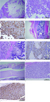
Metastasis was found in 62.3% of the animals. Two weeks after intracardiac injection, metastasis to the brain, spine and femur was detected with bioluminescence imaging with an increasing intensity by week 7. Micro-CT scan confirmed multiple osteolytic lesions at the femur, spine and skull. MRI and the histology were able to show metastasis in the brain and extraskeletal metastasis around the femur.
Figures and Tables
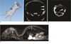
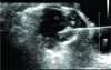 | Fig. 1Ultrasonogram of intracardiac injection to nude mouse. Ultrasound guided intracardiac injection to the left ventricle of nude mouse is shown. The needle tip is seen inside of the left ventricle.
|
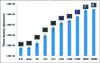 | Fig. 2Bioluminescence photon intensity of serially diluted MDA-MB-231Bo-Luc cell line in 96-well plate.Cells were serially diluted in from 1×10⁵ to 7.8×10³ cells/well. Wells with media (no cells) and distilled water (D.W.) were included as control.
|
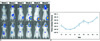 | Fig. 3Growth kinetics of MDA-MB-231Bo-luc cell line in individual mice.A. Bioluminescence images from a representative mice, taken from the dorsal side, with every week intervals after injection of 2 × 10⁵ MDA-MB-231Bo-Luc cells. Brain, femur and spine metastasis was detected.
B. Serial average photon flux intensity from all animal.
|
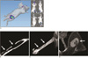 | Fig. 4Multimodal imaging showing femur metastasis.3D (left) and 2D (right small figures in prone, supine, right lateral, and left lateral positions) bioluminescence images, signal was shown on brain and right femur.
Micro-CT image shows cortical osteolysis of the right femur (arrows).
The 3D reconstructed image of micro-CT shows lesion extent of bone metastasis on the femur (arrows).
Axial T2 weighted MR image shows extraskeletal extension of the metastatic tumor (arrow).
|
 | Fig. 5Multimodal imaging showing spine and brain metastasis.3D bioluminescence image shows brain and spine metastasis.
Micro-CT image shows osteolysis at the left temporal bone (arrows).
Micro-CT image of the spine shows cortical destruction on the spine (arrow).
Sagittal T2 weighted MR image shows high signal intensity intracranial and spinal canal mass lesions (arrows).
|
Acknowledgement
This work was supported by the Korean Research foundation Grant funded by the Korean Government (MOEHRD, Basic Research Promotion Fund) (KRF-2008-331-E00249)
References
1. Yoneda T. Arterial microvascularization and breast cancer colonization in bone. Histol Histopathol. 1997; 12:1145–1149.
2. Yoneda T, Sasaki A, Mundy GR. Osteolytic bone metastasis in breast cancer. Breast Cancer Res Treat. 1994; 32:73–84.
3. Arguello F, Baggs RB, Frantz CN. A murine model of experimental metastasis to bone and bone marrow. Cancer Res. 1988; 48:6876–6881.
4. Guise TA, Yin JJ, Taylor SD, Kumagai Y, Dallas M, Boyce BF, et al. Evidence for a causal role of parathyroid hormone-related protein in the pathogenesis of human breast cancer-mediated osteolysis. J Clin Invest. 1996; 98:1544–1549.
5. Corey E, Quinn JE, Bladou F, Brown LG, Roudier MP, Brown JM, et al. Establishment and characterization of osseous prostate cancer models: intra-tibial injection of human prostate cancer cells. Prostate. 2002; 52:20–33.
6. Wang CY, Chang YW. A model for osseous metastasis of human breast cancer established by intrafemur injection of the mda-mb-435 cells in nude mice. Anticancer Res. 1997; 17:2471–2474.
7. Kjonniksen I, Winderen M, Bruland O, Fodstad O. Validity and usefulness of human tumor models established by intratibial cell inoculation in nude rats. Cancer Res. 1994; 54:1715–1719.
8. Peterschmitt J, Bauerle T, Berger MR. Effect of zoledronic acid and an antibody against bone sialoprotein II on MDA-MB-231(GFP) breast cancer cells in vitro and on osteolytic lesions induced in vivo by this cell line in nude rats. Clin Exp Metastasis. 2007; 24:449–459.
9. Bauerle T, Adwan H, Kiessling F, Hilbig H, Armbruster FP, Berger MR. Characterization of a rat model with site-specific bone metastasis induced by MDA-MB-231 breast cancer cells and its application to the effects of an antibody against bone sialoprotein. Int J Cancer. 2005; 115:177–186.
10. Hsu WK, Virk MS, Feeley BT, Stout DB, Chatziioannou AF, Lieberman JR. Characterization of osteolytic, osteoblastic, and mixed lesions in a prostate cancer mouse model using 18F-FDG and 18F-Fluoride PET/CT. J Nucl Med. 2008; 49:414–421.
11. Jenkins DE, Hornig YS, Oei Y, Dusich J, Purchio T. Bioluminescent human breast cancer cell lines that permit rapid and sensitive in vivo detection of mammary tumors and multiple metastases in immune deficient mice. Breast Cancer Res. 2005; 7:R444–R454.
12. Minn AJ, Kang Y, Serganova I, Gupta GP, Giri DD, Doubrovin M, et al. Distinct organ-specific metastatic potential of individual breast cancer cells and primary tumors. J Clin Invest. 2005; 115:44–55.
13. Wetterwald A, van der Pluijm G, Que I, Sijmons B, Buijs J, Karperien M, et al. Optical imaging of cancer metastasis to bone marrow: a mouse model of minimal residual disease. Am J Pathol. 2002; 160:1143–1153.
14. Song H, Shahverdi K, Huso DL, Wang Y, Fox JJ, Hobbs RF, et al. An immunotolerant HER-2/neu transgenic mouse model of metastatic breast cancer. Clin Cancer Res. 2008; 14:6116–6124.
15. Mouchess ML, Sohara Y, Nelson MD Jr, DeCLerck YA, Moats RA. Multimodal imaging analysis of tumor progression and bone resorption in a murine cancer model. J Comput Assist Tomogr. 2006; 30:525–534.
16. Yoneda T, Williams PJ, Hiraga T, Niewolna M, Nishimura R. A bone-seeking clone exhibits different biological properties from the MDA-MB-231 parental human breast cancer cells and a brainseeking clone in vivo and in vitro. J Bone Miner Res. 2001; 16:1486–1495.
17. Jonkers J, Derksen PW. Modeling metastatic breast cancer in mice. J Mammary Gland Biol Neoplasia. 2007; 12:191–203.
18. Hortobagyi GN. Developments in chemotherapy of breast cancer. Cancer. 2000; 88:12 Suppl. 3073–3079.
19. Liotta LA, Kohn EC. The microenvironment of the tumour-host interface. Nature. 2001; 411:375–379.
20. Kim S. Animal models of cancer in the head and neck region. Clin Exp Otorhinolaryngol. 2009; 2:55–60.
21. Rosol TJ, Tannehill-Gregg SH, Corn S, Schneider A, McCauley LK. Animal models of bone metastasis. Cancer Treat Res. 2004; 118:47–81.
22. Sano D, Myers JN. Xenograft models of head and neck cancers. Head Neck Oncol. 2009; 1:32.
23. Rozel S, Galban CJ, Nicolay K, Lee KC, Sud S, Neeley C, et al. Synergy between anti-CCL2 and docetaxel as determined by DWMRI in a metastatic bone cancer model. J Cell Biochem. 2009; 107:58–64.
24. Song HT, Jordan EK, Lewis BK, Liu W, Ganjei J, Klaunberg B, et al. Rat model of metastatic breast cancer monitored by MRI at 3 tesla and bioluminescence imaging with histological correlation. J Transl Med. 2009; 7:88.
 XML Download