

 PDF
PDF ePub
ePub Citation
 Print
Print


Abstract
Purpose
The aim of this study was to investigate ultrasound (US) findings of eggshell calcified thyroid nodules associated with thyroid malignancy and the diagnostic usefulness for US-guided fine-needle aspiration cytology (US-FNAC) of eggshell calcified thyroid nodules.
Materials and Methods
We analyzed 36 eggshell calcified thyroid nodules in 35 patients who underwent thyroid US and US-FNAC from January to December of 2009. We compared the US findings and US-FNAC results with the pathologic results confirmed by surgery.
Results
Twenty eggshell calcified nodules were surgically removed in 19 patients, from which 8 papillary thyroid carcinomas and 12 hyperplasia nodules were confirmed. The sensitivity, specificity, positive, and negative predictive values, as well as accuracy for US diagnosis and US-FNAC of eggshell calcified nodules were 100% and 20%, 25% and 100%, 43.8% and 100%, 100% and 63.6%, and 55% and 66.7%, respectively.
Figures and Tables
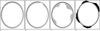 | Fig. 1Schematic drawings of eggshell calcified nodules (Inner thick rim and outer thin rim are an eggshell calcification and a hypoechoic rim, respectively).A. A simple eggshell calcification. B. A disrupted eggshell calcification. C. A thickening of eggshell calcification. D. An eggshell calcified nodule with peripheral thick hypoechoic rim.
|
 | Fig. 2A. A simple eggshell calcification in a 50-year-old woman. Transverse sonogram shows a simple eggshell calcified nodule in right thyroid lobe, and it was pathologically confirmed as nodular hyperplasia because of coexisting thyroid malignancy although it had been cytologically diagnosed as benign after US-FNAC.B. A disrupted eggshell calcification in a 40-year-old woman. Transverse sonogram shows an eggshell calcified nodule with focal defect (arrow) in right thyroid lobe, and it was surgically resected because of suspicious follicular neoplasm in US-FNAC (nodular hyperplasia by pathology).
C. A peripheral thick hypoechoic rim and disrupted eggshell in a 45-year-old woman. Transverse sonogram shows an eggshell calcified nodule with peripheral thick hypoechoic rim (arrows) and disrupted eggshell in left thyroid lobe, and it was pathologically confirmed as papillary thyroid carcinoma because of suspicious cytology.
|
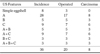
Table 1
Incidence and Pathology Results on the US Features of Eggshell Calcified Thyroid Nodules
Note.─ A-disrupted eggshell calcification, B-peripheral thick hypoechoic rim, C-thickening of eggshell
![]()
References
1. Kwak MS, Baek JH, Kim YS, Jeong HJ. Patterns and Significance of Peripheral Calcifications of Thyroid Tumors seen on Ultrasound. J Korean Radiol Soc. 2005; 53:401–405.
2. Yoon DY, Lee JW, Chang SK, Choi CS, Yun EJ, Seo YL, et al. Peripheral calcification in thyroid nodules: ultrasonographic features and prediction of malignancy. J Ultrasound Med. 2007; 26:1349–1355.
3. Kim BM, Kim MJ, Kim EK, Kwak JY, Hong SW, Son EJ, et al. Sonographic differentiation of thyroid nodules with eggshell calcifications. J Ultrasound Med. 2008; 27:1425–1430.
4. Park M, Shin JH, Han BK, Ko EY, Hwang HS, Kang SS, et al. Sonography of thyroid nodules with peripheral calcifications. J Clin Ultrasound. 2009; 37:324–328.
5. Wang N, Xu Y, Ge C, Guo R, Guo K. Association of sonographically detected calcification with thyroid carcinoma. Head Neck. 2006; 12:1077–1083.
6. Choi JO, Lee JY, Chung K, Choi G. Patterns of Calcification in Thyroid Nodules: Significance and Malignanat Potnetiality. Korean J Head Neck Oncol. 1997; 13:30–34.
7. Khoo ML, Asa SL, Witterick IJ, Freeman JL. Thyroid calcification and its association with thyroid carcinoma. Head Neck. 2002; 24:651–655.
8. Yang YS, Lim HS, Kim YW, Oh JK, Hong KH. The Relative Risk of Cancer in Sonographically Detected Thyroid Nodules with Calcifications. Korean J Otolaryngol-Head Neck Surg. 2004; 47:457–461.
9. Seibering KA, Dutra JC, Grant T, Bajramovic S. Role of intrathyroidal calcifications detected on ultrasound as a marker of malignancy. Laryngoscope. 2004; 114:1753–1757.
10. Kakkos SK, Scopa CD, Chalmoukis AK, Karachalios DA, Spiliotis JD, Harkoftakis JG, et al. Relative risk of cancer in sonographically detected thyroid nodules with calcifications. J Clin Ultrasound. 2000; 28:347–352.
11. Koike E, Noguchi S, Yamashita H, Murakami T, Ohshima A, Kawamoto H, et al. Ultrasonographic characteristics of thyroid nodules: prediction of malignancy. Arch Surg. 2001; 136:334–337.
12. Kim EK, Park CS, Chung WY, Oh KK, Kim DI, Lee JT, et al. New sonographic criteria for recommending fine-needle aspiration biopsy of nonpalpable solid nodules of the thyroid. AJR Am J Roentgenol. 2002; 178:687–691.
13. Frates MC, Benson CB, Charboneau JW, Cibas ES, Clark OH, Coleman BG, et al. Management of thyroid nodules detected at US: society of radiologists in ultrasound consensus conference statement. Radiology. 2005; 237:794–800.
14. Moon WJ, Baek JH, Jung SL, Kim DW, Kim EK, Kim JY, et al. Ultrasonography and ultrasound-based management of thyroid nodules: consensus statement and recommendations. Korean J Radiol. 2011; 12:1–14.
 XML Download