Abstract
Desmoplastic fibroma of bone is a rare benign primary bone tumor that histologically resembles the extra-abdominal desmoid tumor of soft tissues. It is a nonmetastasizing, but locally aggressive tumor that is similar to a desmoid tumor of the soft tissues, and so it is considered "semimalignant". According to a previous report on a series of bone tumors, the incidence rate of desmoplastic fibroma was 0.1-0.3%. Its rarity results in radiologists having a tendency of overlooking the possibility of desmoplastic fibroma of bone during the imaging readings. We report on the imaging findings of desmoplastic fibroma of bone with a review of the relevant literature.
Desmoplastic fibroma of bone is a rare benign primary bone tumor. It was first described in 1958 by Jaffe as a distinct entity from other intraosseous fibrous tumors (1). Desmoplastic fibroma of bone is now considered to be the intraosseous counterpart of the soft tissue desmoid or fibromatosis (2). Recognition of this entity is important because it may be histologically and radiologically confused with benign fibrous lesions or more significantly, with spindle cell sarcomas (3). In the previously published major series of bone tumors, the reported incidence of desmoplastic fibroma was 0.1-0.3% (24). Similar to soft tissue desmoid, desmoplastic fibroma of bone is locally aggressive by nature with a chance for local recurrence, even after treatment (3). We experienced a pathologically confirmed case of desmoplastic fibroma in bone, which locally recurred after treatment. Here, we report on the imaging findings of desmoplastic fibroma of bone and a review of the relevant literature.
In February 2003, a 36-year-old woman presented with right knee pain and swelling and had been referred to our hospital for further evaluation. The physical examination showed a palpable mass with local tenderness on the right proximal tibial area. Her past medical history and laboratory examinations were all unremarkable.
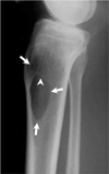
Simple radiographs at that time of diagnosis revealed a well demarcated, oval shaped osteolytic lesion in the proximal metadiaphysis of the tibia. This lesion was eccentrically located in the medullary cavity. The surrounding cortex was intact and no distinctive periosteal reaction was seen. Further, there was a lack of a soft tissue extension (Fig. 1) and the lesion showed internal bony ridges with a narrow transition zone and no matrix mineralization.
Magnetic resonance (MR) imaging was performed si multaneously with simple radiographs. It showed an isointense lesion compared to the adjacent normal muscle on the T1-weighted image and heterogeneous low signal intensity on the T2-weighted image in the tibial metadiaphysis. In contrast to simple radiographs, MRI clearly showed the finding of cortical destruction with soft tissue extension (Figs. 2A-C). The mass showed heterogeneous enhancement on the gadolinium-enhanced images. The hypointense area on T2 weighted image showed less enhancement than the hyperintense area (Fig. 2C).
An incisional biopsy was performed, which resulted in a non-specific fibrosis. The mass underwent complete curettage, and then histologically confirmed as desmoplastic fibroma of bone. The mass was poorly demarcated and infiltrated into the surrounding skeletal muscle tissue. The histologic specimen was identical to the soft tissue desmoid (Fig. 2D). After complete curettage, the patient was discharged without any complication.
About 7 years later, she revisited the hospital with recurring right knee pain for one month. MRI showed a heterogeneous signal intensity mass in the lateral tibial metadiaphysis, which extended into the epiphysis. A large cystic cavity was seen in the tibial metadiaphysis adjacent to the recurred mass, and was considered to be the result of the previous curettage. On the proton density-weighted fat suppressed sequence, the recurrent mass generally showed a hyperintense signal that encompassed the internal areas and there were foci with the T2 low signal (Fig. 3A).
A wide resection was performed and the mass was confirmed to be desmoplastic fibroma. The mass showed histologic features that were basically similar to those of the previous specimen, and the mass was considered to be a recurrence of desmoplastic fibroma of bone. The recurrent mass had more cellular areas and a lesser amount of collagen bundles when compared to the previous specimen (Fig. 3B).
Desmoplastic fibroma of bone is a rare primary tumor of bone. Approximately, 180 cases have been reported in the English medical literature, most of which were case reports. The incidence of desmoplastic fibroma is reported to be 0.1-0.3% of all primary bone tumors (24). Because of its rarity, desmoplastic fibroma of bone has been described in small series in orthopedic, pathologic, and radiologic literatures.
Although desmoplastic fibroma of bone can occur in any age group, most patients tend to be in the third to fourth decades of life (356); the gender distribution of this tumor has not been definitely established. Inwards et al. (3) reported a male predominance, whereas Crim et al. (5) did not find a gender predilection.
Desmoplastic fibroma of bone has been reported in almost all the bones of the skeleton and these lesions occur with a similar frequency in flat and long bones (5). The mandible (22%), femur (15%), pelvic bones (13%), radius (12%) and tibia (9%) are often involved sites (7).
The clinical manifestations are nonspecific and similar to those of other bone tumors. Pain, a palpable mass and/or swelling are the common manifestations. However, some patients have no symptoms and the tumor is discovered incidentally on the plain. About 10-15% of the patients present with a pathologic fracture (356). Histologically, desmoplastic fibroma of bone has identical features to those of the soft tissue desmoid and because of the similar locally aggressive pattern of growth, this tumor is considered as an intraosseous counterpart of the soft tissue desmoids (234). Desmoplastic fibroma is characterized by fibroblasts within a stroma containing various amounts of collagen matrix. The fibroblasts have a uniform spindle shape with ovoid or slender nuclei, and the fibroblasts are evenly distributed in the abundant collagenous fibers that form thick, hyaline strands (356).
The simple radiographic findings of desmoplastic fibroma of bone are a well demarcated, osteolytic tumor that does not contain any mineralized matrix (56). In the long bones, Crim et al. (5) reported that this tumor was oval in shape with its longest dimension aligned with the long axis of the host bone. Further, it usually arises in the metaphysis or metadiaphysis and it may extend into the epiphysis and also into the subchondral bone plate in skeletally mature patients (356). It may also arise in the diaphysis, but only very rare cases (56). The center of the tumor is equally likely to be central or eccentric in the medullary cavity (5). The most consistent radiographic findings for desmoplastic fibroma of bone are as follows: a well defined geographic lesion with a narrow transition zone (96%) and a non-sclerotic margin (94%) as a type 1b geographic lesion with internal pseudotrabeculation (91%) and bone expansion (89%) (5). Investigators from prior studies reported that cortical destruction was seen in 29% of cases and a soft tissue mass was often present. Distinctive periosteal reaction is rare (56). The simple radiographic findings of our case correspond to the findings reported in the literatures.
When the plain radiographic appearances of this tumor are totally assessed, desmoplastic fibromas of bone generally have characteristics of benign bone lesion. Therefore, a radiologist ought to differentiate this tumor from various benign bone lesions such as giant cell tumor, fibrous dysplasia, aneurysmal bone cyst, chondromyxoid fibroma, non-ossifying fibroma, and simple bone cyst. Although the radiographic appearance of desmplastic fibroma of bone is somewhat similar to that of other benign lytic bone lesions, some characteristics are useful in the differential diagnosis. A giant cell tumor, like desmoplastic fibroma of bone, may extend into the epiphysis and to the subchondral bone plate from the metaphysic, but it is round rather than oval. Fibrous dysplasia tends to occupy a longer segment of bone, contains a mineralized matrix, and often has a sclerotic rim. Bone expansion in aneurysmal bone cyst tends to have an eccentric, "blow-out" appearance, as opposed to the fusiform expansion usually seen with desemoplastic fibroma. Chondromyxoid fibroma is usually more eccentrically located, with a delicately scalloped border and thin marginal sclerosis. Nonossifying fibroma is distinguished by its eccentricity, scalloped margin, and a sclerotic rim. Pseudotrabeculation in chondromyxoid fibroma and nonossifying fibroma tends to appear more curvilinear than in desmoplastic fibroma. A simple bone cyst may appear identical to desmoplastic fibroma on plain radiographs but often can be distinguished by its fluid attenuation on CT or MR scans (5).
When cortical destruction with soft tissue extension is seen, the appearance of this lesion can be similar to that of malignant bone lesions such as fibrosarcoma and lowgrade osteosarcoma. However, radiographs are useful in the differentiation, usually revealing a permeative pattern of bone destruction and a wider zone of transition in low-grade osteosarcoma, but a geographic pattern in almost all cases of desmplastic fibroma of bone. Lowgrade osteosarcoma, unlike desmoplastic fibroma of bone, is typically associated with areas of mineralized tumor matrix (5).
In contrast to the well-established radiographic appearance, the MR findings of desmoplastic fibroma of bone are scarce. One series in the radiologic literature analyzed and discussed the MR features of this tumor (8). The MR features of desmoplastic fibroma of bone are as follows. On a T1-weighted sequence, the tumor shows an isointense or hypointense signal compared to adjacent normal muscle. Almost all of the cases show large, hypointense or isointense signal areas on the T2-weighted sequence and T2 low signal is the predominant signal pattern. After intravenous gadolinium contrast administration, the lesion shows heterogeneous contrast enhancement with areas of intense enhancement and none to minor enhancement in other areas (89).
Similar to soft tissue desmoid, the areas with hypointense and hyperintense signal on a T2-weighted sequence may correspond to the collagenous parts and the more cellular areas, respectively (910). Heterogeneous enhancement of the lesion seems to reflect the variable composition of the cellular part and the collagenous matrix (9). In our case, the hyperintense area on the T2-weighted image was more intensely enhanced than that of the area of T2 low signal. However, no histologic correlation studies exist for desmoplastic fibroma of bone.
As mentioned earlier in the introduction, desmoplastic fibroma of bone locally aggressive by nature, like its soft tissue counterpart, sometimes resulting in local recurrence. The same as in our case, the patients treated with curettage alone show a high rate of recurrence. Therefore, wide resection is the treatment of choice, and adequate resection can be expected to result in no recurrence (5).
Areas of T2 low signal is an interesting finding of desmoplastic fibroma of bone. However, it has also been described in other bone lesions such as giant cell tumor, fibrous dysplasia, non-ossifying fibroma, lymphoma, and leiomyosarcoma (89). As previously mentioned, although the radiographic features of desmplastic fibroma of bone is an osteolytic bony lesion with a benign appearance, some radiographic findings are helpful to differentiate it from other benign osteolytic bone lesions with T2 low signal on MRI, including giant cell tumor, fibrous dysplasia, and non-ossifying fibroma.
In conclusion, a predominant hypointense signal on MRI is not a characteristic feature of desmoplastic fibroma of bone. However, T2 low signal is rarely encountered in benign radiographic appearing osteolytic bone tumors. The combination of T2 low signal and some radiographic characteristics may help narrow the differential diagnosis, and radiologists ought to consider the possibility of desmoplastic fibroma of bone.
Figures and Tables
Fig. 1
The plain lateral and anteroposterior (not shown) radiographs of the proximal right tibia in a 36-year-old woman show a well marginated, osteolytic lesion (arrows) with internal trabeculation (arrow head) within the proximal tibial metadiaphysis. There is no evidence of matrix mineralization or cortical destruction.

Fig. 2
The coronal T1-weighted images (A) show an isointense lesion compared to the adjacent normal muscle within the proximal tibial metadiaphysis with cortical destruction and a soft tissue extension.
The axial T2-weighted image (B) at a same level shows a hyperintense lesion in the medullary area (asterisk), which is hypointense to isointense relative to the peripheral area (arrow head). Note that the area of T2 low signal is more extensive than the hyperintense medullary area.
The axial T1-wegihted image after gadolinium contrast administration (C) shows heterogeneous enhancement of the lesion. The hyperintense area on the T2-weighted image (asterisk) is more intensely enhanced than the area with T2 low signal (arrow head).
Microscopic examination (D) shows monotonous spindle shaped fibroblasts with slender nuclei interspersed in the hyaline stroma of thick, wavy bands of collagen fiber (H & E, × 200).

Fig. 3
MRI is performed to determine whether there is tumor recurrence. The coronal proton density-weighted fat suppressed image (A) shows a hyperintense lesion with foci of T2 low signal within the lateral proximal tibial metadiaphysis. The cavity of fluid signal intensity in the medulla (asterisk) seems to be the result of previous curettage. In contrast to the previous lesion in Fig. 2A, this lesion extends into the epiphysis (arrow). Note that the overall signal intensity of the lesion is higher than that of the previous lesion in Fig. 2B.

References
1. Jaffe HL. Tumors and tumorous conditions of the bones and joints. Philadelphia: Lea & Febiger;1958. p. 298–303.
2. Fornasico V, Pritzker KPH, Bridge JA. Pathology and genetics of tumours of soft tissue and bone. In : Fletcher CDM, Unni KK, Merten SF, editors. Pathology and genetics of tumours of soft tissue and bone. Lyon: IARC Press;2002. p. 288.
3. Inwards CY, Unni KK, Beabout JW, Sim FH. Desmoplastic fibroma of bone. Cancer. 1991; 68:1978–1983.
4. Mirra JM, Picci P, Gold RH. Bone tumors: clinical, radiologic, and pathologic correlations. Philadelphia: Lea & Febiger;1989. p. 735–747.
5. Crim JR, Gold RH, Mirra JM, Eckardt JJ, Bassett LW. Desmoplastic fibroma of bone: radiographic analysis. Radiology. 1989; 172:827–832.
6. Taconis WK, Schutte HE, van der Heul RO. Desmoplastic fibroma of bone: a reported of 18 cases. Skeletal Radiol. 1994; 23:283–288.
7. Bohm P, Krober S, Greschniok A, Laniado M, Kaiserling E. Desmoplastic fibroma of the bone. A reported of two patients, review of the literature, and therapeutic implications. Cancer. 1996; 78:1011–1023.
8. Frick MA, Sundaram M, Unni KK, Inwards CY, Fabbri N, Tretani F, et al. Imaging findings in desmoplastic fibroma of bone: distinctive T2 chracteristic. AJR Am J Roentgenol. 2005; 184:1762–1767.
9. Vanhoenacker FM, Hauben E, De Beuckeleer LH, Willemen D, Van Marck E, De Schepper AM. Desmoplastic fibroma of bone: MRI features. Skeletal Radiol. 2000; 29:171–175.
10. Sundaram M, McGuire MH, Schajowicz F. Soft-tissue masses: histologic basis for decreased signal (short T2) on T2-weighted MR images. AJR Am J Roentgenol. 1987; 148:1247–1767.


 PDF
PDF ePub
ePub Citation
 Print
Print


 XML Download