

 PDF
PDF ePub
ePub Citation
 Print
Print


Abstract
Purpose
To evaluate the reproducibility of 3-dimensional histomorphometry for the microarchitecture analysis of trabecular bone parameters using multidetector computed tomography (MDCT).
Materials and Methods
Thirty-six specimens from porcine vertebral bodies were imaged five times with a 64-detector row MDCT system using the same scan protocols. Locations of the specimens were nearly identical through the scans. Three-dimensional structural parameters of trabecular bone were derived from the five data sets using image analyzing software. The features measured by the analysis programs were trabecular bone volume, trabecular bone volume/tissue volume, trabecular thickness, trabecular separation, trabecular number, trabecular bone pattern factor, structural model index.
Results
The structural trabecular parameters showed excellent reproducibility through repeated scanning. Intraclass correlation coefficients of all seven structural parameters were in the range of 0.998 to 1.000. Coefficients of variation of the six structural parameters, excluding structural model index, were not over 1.6%.
Figures and Tables
Fig. 1
Bland-Altman plots of paired results of the three-dimensional structural parameters between the maximum value and minimum value.
Note.─ BV= trabecular bone volume, BVTV= trabecular bone volume/tissue volume, Tb_Th= trabecular thickness, Tb_Sp= trabecular separation, Tb_N= trabecular number, Tb_Pf= trabecular bone pattern factor, SMI= structural model index
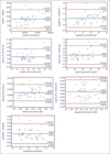
Fig. 2
Dispersion of standard deviation versus mean and coefficient of variation versus mean.
Note.─ BV= trabecular bone volume, BVTV= trabecular bone volume/tissue volume, Tb_Th= trabecular thickness, Tb_Sp= trabecular separation, Tb_N= trabecular number, Tb_Pf= trabecular bone pattern factor, SMI= structural model index, SD= standard deviation, CV= coefficient of variation
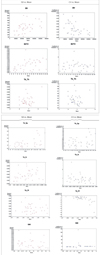
Table 2
Distribution of Coefficients of Variation for the Structural Parameters Repeatedly Scanned Five Times
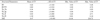
Note.─ BV= trabecular bone volume, BV/TV= trabecular bone volume/tissue volume, Tb_Th= trabecular thickness, Tb_Sp= trabecular separation, Tb_N= trabecular number, Tb_Pf= trabecular bone pattern factor, SMI= structural model index, SD= standard deviation, CV= coefficient of variation, Min. value= Minimum value, Max. value= Maximum value
References
1. Carballido-Gamio J, Majumdar S. Clinical utility of microarchitecture measurements of trabecular bone. Curr Osteoporos Rep. 2006; 4:64–70.
2. Patel PV, Prevrhal S, Bauer JS, Phan C, Eckstein F, Lochmuller EM, et al. Trabecular bone structure obtained from multislice spiral computed tomography of the calcaneus predicts osteoporotic vertebral deformities. J Comput Assist Tomogr. 2005; 29:246–253.
3. Keaveny TM, Morgan EF, Niebur GL, Yeh OC. Biomechanics of trabecular bone. Annu Rev Biomed Eng. 2001; 3:307–333.
4. Chappard D, Retailleau-Gaborit N, Legrand E, Basle MF, Audran M. Comparison insight bone measurements by histomorphometry and microCT. J Bone Miner Res. 2005; 20:1177–1184.
5. Chappard D, Guggenbuhl P, Legrand E, Basle MF, Audran M. Texture analysis of X-ray radiographs is correlated with bone histomorphometry. J Bone Miner Metab. 2005; 23:24–29.
6. Boutry N, Cortet B, Dubois P, Marchandise X, Cotten A. Trabecular bone structure of the calcaneus: preliminary in vivo MR imaging assessment in men with osteoporosis. Radiology. 2003; 227:708–717.
7. Krug R, Carballido-Gamio J, Burghardt A, Haase S, Sedat J, Moss W, et al. Wavelet Based Characterization of vertebral trabecular bone structure from MR images of specimen at 3 tesla compared to microCT measurements. Conf Proc IEEE Eng Med Biol Soc. 2005; 7:7040–7043.
8. Lasbleiz J, Burgun A, Marin F, Rolland Y, Duvauferrier R. Vertebral trabecular network analysis on CT images. J Radiol. 2005; 86:645–649.
9. Cortet B, Chappard D, Boutry N, Dubois P, Cotten A, Marchandise X. Relationship between computed tomographic image analysis and histomorphometry for microarchitectural characterization of human calcaneus. Calcif Tissue Int. 2004; 75:23–31.
10. Link TM, Vieth V, Stehling C, Lotter A, Beer A, Newitt D, et al. High-resolution MRI vs multislice spiral CT: which technique depicts the trabecular bone structure best? Eur Radiol. 2003; 13:663–671.
11. Torres A, Lorenzo V, Gonzalez-Posada JM. Comparison of histomorphometry and computerized tomography of the spine in quantitating trabecular bone in renal osteodystrophy. Nephron. 1986; 44:282–287.
12. Genant HK, Delmas PD, Chen P, Jiang Y, Eriksen EF, Dalsky GP, et al. Severity of vertebral fracture reflects deterioration of bone microarchitecture. Osteoporos Int. 2007; 18:69–76.
13. Jiang SD, Jiang LS, Dai LY. Spinal cord injury causes more damage to bone mass, bone structure, biomechanical properties and bone metabolism than sciatic neurectomy in young rats. Osteoporos Int. 2006; 17:1552–1561.
14. Gustafsson BI, Westbroek I, Waarsing JH, Waldum H, Solligard E, Brunsvik A, et al. Long-term serotonin administration leads to higher bone mineral density, affects bone architecture, and leads to higher femoral bone stiffness in rats. J Cell Biochem. 2006; 97:1283–1291.
15. Byers BA, Guldberg RE, Hutmacher DW, Garcia AJ. Effects of Runx2 genetic engineering and in vitro maturation of tissue-engineered constructs on the repair of critical size bone defects. J Biomed Mater Res A. 2006; 76:646–655.
16. Trisi P, Rebaudi A, Calvari F, Lazzara RJ. Sinus graft with biogran, autogenous bone, and PRP: a report of three cases with histology and micro-CT. Int J Periodontics Restorative Dent. 2006; 26:113–125.
17. Vigorita VJ. The bone biopsy protocol for evaluating osteoporosis and osteomalacia. Am J Surg Pathol. 1984; 8:925–930.
18. Link TM, Majumdar S, Grampp S, Guglielmi G, van Kuijk C, Imhof H, et al. Imaging of trabecular bone structure in osteoporosis. Eur Radiol. 1999; 9:1781–1788.
19. Hildebrand T, Ruegsegger P. A new method for the model-independent assessment of thickness in three-dimensional images. J Microsc. 1997; 185:67–75.
20. Hahn M, Vogel M, Pompesius-Kempa M, Delling G. Trabecular bone pattern factor—a new parameter for simple quantification of bone microarchitecture. Bone. 1992; 13:327–330.
21. Kleerekoper M, Villanueva AR, Stanciu J, Rao DS, Parfitt AM. The role of three-dimensional trabecular microstructure in the pathogenesis of vertebral compression fractures. Calcif Tissue Int. 1985; 37:594–597.
22. Ross PD, Wasnich RD, Davis JW. Fracture prediction models for osteoporosis prevention. Bone. 1990; 11:327–331.
23. Ross PD, Davis JW, Vogel JM, Wasnich RD. A critical review of bone mass and the risk of fractures in osteoporosis. Calcif Tissue Int. 1990; 46:149–161.
24. Link TM, Vieth V, Matheis J, Newitt D, Lu Y, Rummeny EJ, et al. Bone structure of the distal radius and the calcaneus vs BMD of the spine and proximal femur in the prediction of osteoporotic spine fractures. Eur Radiol. 2002; 12:401–408.
25. Wehrli FW, Song HK, Saha PK, Wright AC. Quantitative MRI for the assessment of bone structure and function. NMR Biomed. 2006; 19:731–764.
26. Boutroy S, Bouxsein ML, Munoz F, Delmas PD. In vivo assessment of trabecular bone microarchitecture by high-resolution peripheral quantitative computed tomography. J Clin Endocrinol Metab. 2005; 90:6508–6515.
27. Chen P, Miller PD, Recker R, Resch H, Rana A, Pavo I, et al. Increases in BMD correlate with improvements in bone microarchitecture with teriparatide tretment in postmenopausal women with osteoporosis. J Bone Miner Res. 2007; 22:1173–1180.
28. Benhamou CL. Effects of osteoporosis medications on bone quality. Joint Bone Spine. 2007; 74:39–47.
29. Bredella MA, Misra M, Miller KK, Madisch I, Sarwar A, Cheung A, et al. Distal radius in adolescent girls with anorexia nervosa: trabecular structure analysis with high-resolution flat-panel volume CT. Radiology. 2008; 249:938–946.
 XML Download