

 PDF
PDF ePub
ePub Citation
 Print
Print


Neuroendocrine tumors arise from enterochromaffin cells which are widely distributed in the body. The sites most often affected by these tumors, in order of importance, are the gastrointestinal and bronchopulmonary tracts (1). However, primary neuroendocrine tumors in the presacral region are extremely rare. Even though there were a few reports about neuroendocrine tumor of the presacral region, to the best of our knowledge, primary malignant neuroendocrine tumor of the presacral region has not been described with a focus on the imaging findings. Therefore, we report an extremely rare case of primary well-differentiated neuroendocrine carcinoma of the presacral region.
Case Report
A 65-year-old man visited the hospital with severe coccyx pain during sleep. The patient was previously healthy with no history of altered bowel habits, rectal bleeding, or urinary symptoms. A general physical examination was unremarkable. A digital rectal examination revealed a firm tender lesion in the retrorectal space 5-8 cm above the anal verge. Perianal examination was normal. The rectal mucosa was normal at sigmoidoscopy. Values for tumor markers, including the carcinoembryonic antigen and CA 19-9, were within normal ranges.
Multi-detector computed tomography (MDCT) was performed to evaluate presacral and coccygeal mass. In addition, we performed pelvic magnetic resonance imaging (MRI) to assess the extent of the lesion as well as the associated findings.
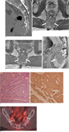
MDCT revealed a well-defined, lobulated, homogeneous 4 cm mass in the presacral area. The mass was located between the rectum and coccyx, and showed soft tissue attenuation with tiny calcifications at its upper portion. The mass tightly abutted the coccyx with resultant pressure erosion (Fig. 1A). On the pelvic MRI, it was difficult to identify the fat plane between the mass, coccyx, and levator ani muscle. However, fat planes between the rectum and mass were well preserved. The lesion showed isointensity on the T1-weighted images and slight hyperintensity on the T2-weighted images. Multiple hypointense septa-like structures within the mass and eccentric focal hyperintense foci at the inferior aspect of the mass were seen on T1 and T2-weighted images (Figs. 1B-D).
A surgical finding revealed a 4.5 × 4.3 × 3.5 cm presacral mass with focal invasion of the coccyx. The lesion was easily separated from the rectum. The presacral mass was resected with removal of the invaded coccyx. After surgery, the patient received radiation therapy because of positive resection margin for tumor invasion.
The gross specimen consisted of a relatively well-defined, solid mass measuring 4.5 × 4.3 × 3.5 cm in size with an attached 2 × 1.7 cm bone. A cut section of the gross specimen showed a homogeneous yellowish color, multiple foci of necrosis, and calcification. With light microscopy, tumor cells were found to be composed of round or fusiform cells arranged in sheets, nests, and rosette-like structure with multiple tumor emboli in lymphovascular spaces and 2-7 mitotic count/10HPF (Fig. 1E). With immunohistochemical staining, the tumor cells were strongly positive for synaptophysin, EMA, and c-kit (Fig. 1F). These findings were consistent with well differentiated neuroendocrine carcinoma. There was no associated lesion such as a tailgut cyst or cystic hamartoma.
On the follow-up abdominal MDCT and PET/CT after surgical excision of the mass, metastases in both internal iliac lymph nodes were detected, but there was no other abnormality suggesting the presence of other primary tumor or metastases (Fig. 1G).
Discussion
Neuroendocrine tumors, derived from endocrine cells of the gastrointestinal tract, are potentially malignant tumors capable of hormonal activity. According to the recent World Health Organization classification, these tumors are classified into three categories; well-differentiated neuroendocrine tumor (carcinoids), well-differentiated neuroendocrine carcinoma (malignant carcinoids), and poorly differentiated neuroendocrine carcinoma. Each category corresponds to a different prognosis, clinical behavior, and treatment possibilities, regardless of the degree of differentiation and site of the primary tumor (2).
Histologically, neurosecretory granules often demonstrate an affinity for soluble silver salts and are confirmed on electron microscopy. Immunohistochemical staining is positive for chromogranin, synaptophysin, and neuron-specific enolase (NSE) (2). The most common finding on microscopic examination is the trabecular growth pattern which is associated with rectal carcinoid tumors (3).
The vast majority of neuroendocrine tumors are found in the gastrointestinal tract and tend to most commonly arise in the small intestine (45%), followed by the rectum (20%), appendix (10%), colon (11%), and stomach (7%) (1).
The presacral region is defined as the potential space delineated by the rectum anteriorly, the sacrum posteriorly, the peritoneal reflection superiorly, and the perineal muscles inferiorly (4). Various tumors or tumorlike lesions including teratoma, chordoma, myxopapillary ependymoma, paraganglioma, schwannoma, liposarcoma, tailgut cyst, and metastatic tumor can occur in this area with increased incidence. However, their radiologic findings may usually be nonspecific for differential diagnosis.
Among these diseases, teratoma and liposarcoma are relatively well differentiated because of fat component. In case of tail gut cyst, it usually shows fluid signal intensity and no enhancement on MRI. In our case, the radiologic finding of the presacral mass was suggested the presence of having chordoma because of tumor location, erosion of the coccyx, and calcifications. We could exclude the possibility of metastatic tumor because there was no other primary tumor detected at PET/CT or follow-up abdominal MDCT.
Only 19 cases of involving the presacral space have been reported in the world literature. Neuroendocrine tumors appear to occur more frequently in females than in males; most of them showed benign behavior with only four cases proven to be malignant with a recurrence at the primary site or distant metastatic dissemination (4567). They represent the direct extension or metastatic spread from rectal primary tumors in most cases (3). In our case, however, the fat plane between the rectum and mass was well preserved on the MRI, and there was no abnomarlity in the rectum on sigmoidoscopy. On surgical exploration, the mass was easily separated from the rectum. In addition, there was no primary neuroendocrine tumor of other sites at follow-up PET/CT or abdominal MDCT. Therefore, we thought it to be primary neuroendocrine tumor arising from the presacral space rather than malignancy arising from the rectum or metastasis from neuroendocrine tumor of other sites. Moreover, in our case, two large metastases in both internal iliac lymph nodes representing its malignant behavior, was developed after surgery of primary tumor.
In a review of the literature, presacral neuroendocrine carcinoma was associated with some anomalies such as tailgut cysts, sacrococcygeal teratoma, and imperforated anus (8). In our case, despite the absence of an underlying anomaly such as tailgut cyst or teratoma, development as a result of the tumor from the rest of the hindgut may be hypothesized. Horenstein et al. (4) reported that neuroendocrine tumors of the presacral space are histopathologically similar to rectal neuroendocrine tumors. This theory is that the presence of remnant hindgut cells may be the basis for the development of neuroendocrine tumors in this region, whether associated with tailgut cysts or not.
In conclusion, we report an extremely rare case of primary neuroendocrine carcinoma of the presacral region focused on imaging findings. Although its radiologic features are nonspecific, primary neuroendocrine carcionoma should be considered in the differential diagnosis of presacral soft tissue masses.
 XML Download