

 PDF
PDF ePub
ePub Citation
 Print
Print


INTRODUCTION
The petrous apex is the portion of the temporal bone, lying anteromedial to the inner ear. It is located between the sphenoid bone and the occipital bone and terminates at the foramen lacerum. This area cannot be directly examined, so lesions of the petrous apex represent a challenging diagnostic and therapeutic problem to radiologists or neurotologists, even though they are the most experienced in dealing with these types of lesions. CT or MRI plays a primary role in the evaluation of lesions located in this area.
The petrous apex can be involved with various cystic and solid lesions (12). Cystic lesions, such as cholesterol granuloma, mucocele, and congenital or acquired cholesteatoma, are much more common than solid lesions (3). A petrous apex cephalocele (PAC) is a rare lesion that arises from Meckel's cave, secondarily erodes the petrous apex, and is characterized by residing in an eccentric location, having a cystic appearance, and is contiguous to the posterolateral aspect of Meckel's cave on imaging findings (34).
Recently, we experienced two cases of PAC, in which the temporal bone or brain CT and MRI including diffusion-weighted sequence demonstrated typical imaging findings at the bilateral petrous apex. In this study, we describe the imaging findings of CT and MRI with a review of the literature.
Case Reports
Case 1
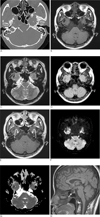
A 42-year-old woman presented with progressive bilateral tinnitus and mild hearing impairment. The leftsided tinnitus developed 20 years ago, but she was not treated at that time. The right-sided tinnitus and mild hearing impairment recently developed. A physical examination revealed that the tinnitus was of venous origin. Initially, a temporal bone CT was obtained and revealed sharply demarcated, multilobulating erosive lesions with a thin cortical bone outline at the bilateral petrous apex (Fig. 1A). MRI demonstrated multilobulating cystic lesions, contiguous to Meckel's cave, at the bilateral petrous apex on T1-weighted, T2-weighted, and a fluid-attenuated inversion recovery (FLAIR) image (Figs. 1B-D). The signal intensity was the same as cerebrospinal fluid (CSF), and partial thin rim enhancement was noted on contrast-enhanced T1-weighted image (Fig. 1E), and diffusion-weighted image (DWI). Moreover, an apparent diffusion coefficient (ADC) map confirmed the CSF-like content of the lesions (Figs. 1F, G). The characteristic findings on CT and MRI suggested a PAC, so she did not opt for surgery and was instead underwent conservative treatment. The patient was then admitted 6 years ago due to amenorrhea and galactorrhea. A sellar MRI at that time revealed empty sella (Fig. 1H).
Case 2
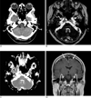
A 63-year-old woman presented with a headache and progressive left leg weakness for 2 weeks. She also complained of right hemifacial hemispasm for 10 years. A brain CT showed cystic lesions at bilateral petrous apex (Fig. 2A). Brain MRI demonstrated multilobulating cystic lesions at the bilateral petrous apex, which were isointense to CSF and contiguous to Meckel's cave. The lesions encroached to anterior wall of internal auditory canal and were located at the superior aspect of carotid canals (Fig. 2B). A DWI and ADC map confirmed the CSF-like content of the lesions (Fig. 2C). The lesions were not enhanced on contrast-enhanced T1-weighted image, but localized meningeal enhancement was noted at high frontal lobes along the midline (Fig. 2D). Viral meningitis was detected by CSF tapping, so antiviral drugs were administered and symptoms were improved. On MRI taken after 17 months, previous meningeal enhancement disappeared and the lesions at the bilateral petrous apex were not changed in size and shape. Coronal T1-weighted image showed flattening of the pituitary gland, suggesting empty sella (not shown here).
DISCUSSION
Petrous apex has a complex relationship with adjacent brain, cranial nerves, and major vessels, and it cannot be examined directly. So, radiologic imaging by CT or MRI plays a primary role in the evaluation of lesions in this area. Petrous apex lesions consist of normal variations, cystic lesions, and solid lesions (125). When there is a cystic lesion at the petrous apex, retained fluid, benign obstructive lesions of air cells (cholesterol granuloma, mucocele), congenital or acquired cholesteatoma, and apical petrositis can be considered.
Trigeminal nerve courses from the prepontine cistern into Meckel's cave over the petrous apex. The porus trigeminus passes through the trigeminal nerve and enters the trigeminal cistern. Along the posterior margin of Meckel's cave, there is normal smooth anterior petrous apex scalloping, trigeminal impression. If the intracranial pressure increases, herniation of the meninges and CSF can cause erosion of this area and promote the subsequent development of a PAC. Moreover, the center of a PAC is the posterolateral margin of Meckel's cave, and the cystic lesion of the petrous apex is contiguous with Meckel's cave. Hence, other cystic lesions arise from and expand the petrous apex within. A PAC is also known as Meckel's cave diverticulum, and the pathologic diagnosis of a PAC has varied from meningocele to arachnoid cyst. When the defect is present at dura, only the CSF-containing arachnoid membrane can herniate into the petrous apex (46). But if arachnoid cyst involves the posterior petrous apex, the imaging feature is quite different from the typical findings of PAC (6).
There are 31 patients with PAC described in the literature. PAC can occur unilaterally or bilaterally. Twenty four out of the 31 reported PAC cases were unilateral, and 16 of the 22 cases in which PAC was unilateral and the site was mentioned, involved right side. Moreover, about three-fourths of cases occurred in women (3). In some patients, PAC may cause trigeminal neuropathy or CSF leak in the form of otorrhea or rhinorrhea due to direct communication between PAC and mastoid air cells, middle ear cavity, or Eustachian tube, and require surgical intervention (234567). All cases complicated by a CSF leak occurred in children (3). A CSF leak can cause recurrent meningitis, tinnitus, postural headache, or conductive hearing loss (8). So, the possible relationship of a PAC with tinnitus or hearing impairment of the patient in case 1 cannot be excluded entirely. A PAC could be discovered incidentally and imaged for reasons clearly unrelated to PAC. A careful assessment is needed to avoid unnecessary treatment or surgical intervention.
Solid lesions of the petrous apex can be easily distinguished by the appearance of bone erosion, signal intensity, and enhancement pattern on MRI. Cystic lesions except cholesterol granuloma, which is shown as high signal intensity on T1-weighted and T2-weighted images, have similar signal intensity to a PAC, but FLAIR and DWI can discriminate these lesions. Also, PAC has a characteristic finding such that the cystic lesions are contiguous to Meckel's cave. In our cases, bilateral petrous apex lesions have same signal intensity as CSF on FLAIR and DWI (5). Petrous air-cell effusions (retained fluid) have similar signal intensity to CSF, but it has a preserved air-cell trabeculation and no expansile characteristics (9). A PAC can present as unenhanced or enhanced with thin wall enhancement pattern, and it is different from inflammatory lesions.
Primary empty sella is reported in 5-35% of the general population. The cause of primary empty sella is believed to be a result of impaired CSF absorption, leading to intracranial hypertension and subsequent herniation of the meninges and CSF into the sella, through a defect at the sellar diaphragm (10). It is mechanically similar to CSF expansion and erosion of adjacent bony structures to PAC. The coincidence of a PAC and empty sella can be explained by the same mechanism. In a report by Alorainy (3), all of the five patients with a PAC had variable degrees of empty sella. In our two cases, mild to moderate empty sella was found. So, in spite of the small number of patients, the association of PAC and primary empty sella raise a strong suspicion of a possible etiologic relationship given the similar findings in the literature.
In summary, sharp multilobulating bony erosion with a thin cortical bone outline on CT, eccentric location, cystic appearance, and contiguity to the posterolateral aspect of Meckel's cave are typical findings of PAC. Distinctive imaging findings of PAC allow the radiologist to make this diagnosis with a high degree of certainty, so unnecessary surgical intervention can be avoided.
 XML Download