

 PDF
PDF ePub
ePub Citation
 Print
Print


Abstract
A high-voltage electrical burn caused extensive deep muscle injuries beneath a relatively small skin wound at the contact point. Hidden, undetected deep muscle injuries have a tendency for progressive tissue necrosis, which can lead to major amputations or sepsis. The radiologic features of this rare, sometimes life-threatening injury have occasionally been described in the literature. However, to the best of our knowledge, there have been no reports on a case of pyogenic arthritis of the ankle joint following a high-voltage electrical burn involving the lower extremity. We report a case of the pyogenic arthritis of the ankle joint following a high-voltage electrical burn involving the lower extremity.
Figures and Tables
Fig. 1
A 38-year-old man with high-voltage electrical burn.
Conventional angiography of the left lower extremity shows complete occlusion of distal anterior tibial and dorslis pedis arteries. There is a normal flow of the posterior tibial artery (arrow).
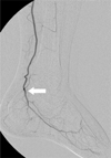
Fig. 2
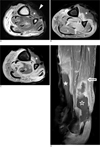
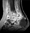
A. Axial T2-weighted (3600/109) turbo spin-echo MR image shows a heterogenous high signal intensity in the entire anterior muscle groups (asterisk) and flexor hallucis longus (FHL) muscle (open asterisk). There is a large anterior defect of the skin and subcutaneous tissue in the distal one-third of the lower leg (arrowhead).
B-D. Gadolinium-enhanced axial (B, C) and sagittal (D) T1-weighted spin-echo MR images obtained with fat saturation show peripheral rim enhancement in the entire anterior muscle groups (asterisks) and part of FHL muscle (open asterisk), representing nonperfused and nonviable areas of edematous muscles. There is diffuse enhancement of FHL muscle (open arrows) at the proximal level, representing perfused and viable area of edematous muscle.
References
1. Fleckenstein JL, Chason DP, Bonte FJ, Parkey RW, Hunt JL, Purdue GF, et al. High-voltage electric injury: assessment of muscle viability with MR imaging and Tc-99m pyrophosphate scintigraphy. Radiology. 1995; 195:205–210.
2. Lee SH, Lee DH, Chang JD, Jun BH. Pyogenic Infection of Deep Joint after Major Burn Injury. J Korean Orthop Assoc. 2005; 40:992–1000.
3. Jang TY, Jo YG, Moon JH, Kim HC, Jo JH. Arteriographic Features in Patients with High-voltage Electric Burn. J Korean Burn Soc. 2007; 10:131–134.
4. Ohashi M, Koizumi J, Hosoda Y, Fujishiro Y, Tuyuki A, Kikuchi K. Correlation between magnetic resonance imaging and histopathology of an amputated forearm after an electrical injury. Burns. 1998; 24:362–368.
5. Choi MN, Lee GK, Suh KJ, Kang IW, Hwang DH, Lee ES, et al. Arteriographic and MR Imaging Findings of a High-Voltage Electrical Burn in the Upper Extremity: A Case Report. J Korean Soc Radiol. 2010; 62:389–392.
6. Moon SY, Kim HC. Investigation of Amputations Related to Electrical Burns. J Korean Burn Soc. 2002; 5:38–56.
7. Song SE, Kim HC, Jang YS, Lee DR. Epidemiologic Analysis of Electric Burns. J Korean Burn Soc. 2007; 10:1–12.
8. Hong SH, Kim SM, Ahn JM, Chung HW, Shin MJ, Kang HS. Tuberculous versus pyogenic arthritis: MR imaging evaluation. Radiology. 2001; 218:848–853.
9. Karchevsky M, Schweitzer ME, Morrison WB, Parellada JA. MRI findings of septic arthritis and associated osteomyelitis in adults. AJR Am J Roentgenol. 2004; 182:119–122.
10. Choi YY. Nuclear Medicine Imaging Diagnosis in Infectious Bone Diseases. Nucl Med Mol Imaging. 2006; 40:193–199.
 XML Download