

 PDF
PDF ePub
ePub Citation
 Print
Print


Although the roles of endoscopic retrograde biliary procedures and computed tomography / magnetic resonance imaging have increased, percutaneous biliary procedures including PTC, PTBD, and percutaneous stent placement remains significant in the evaluation and treatment of biliary obstructive diseases, especially for biliary hilar obstruction (123).
PTBD has been regarded as a drainage-first procedure. Thus, a duct puncture is commonly followed by the insertion of an introducing system to deliver a 0.035 inch wire followed by a drainage catheter. However, during PTBD, it is not uncommon to encounter situations that require manipulations in the bile duct, which cannot be achieved with an introducing system only. For instance, when evaluating an obstructing lesion below the obstructing segment, when an approach to most appropriate site for effective biliary drainage fails, when crossing a tight stricture or an obstructing segment, and when aspirating retained bile before the drainage catheter insertion (345678).
To overcome these problems, we designed a catheter dedicated to percutaneous biliary intervention which have a short stiff shaft and an angled tip for good torque and direction control, as well as terminal side holes for aspiration. We aimed to evaluate the usefulness of this catheter in facilitating the representative percutaneous biliary interventional procedure; PTBD.
MATERIALS AND METHODS
Catheter
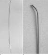
The manipulation catheter (Park's bright; A & A, Seongnam, Korea) has a short (40 cm in length) and stiff-braided shaft made of polyurethane. The terminal 1 cm segment is tapered with a 45° angle (Fig. 1). The stiff shaft and tip make it possible to insert the catheter through the 0.035 inch guide wire into the liver. The angulated tip facilitates direction control and negotiation of the obstructed segment. The catheter has two side holes in the terminal segment to facilitate the aspiration of bile during manipulation.
Patients
Between July 2007 and December 2007, 105 consecutive patients underwent an initial PTBD procedure in our angiographic suite. The catheter was used in all PTBD procedures unless unnecessary or harmful. Thus, 14 of 105 patients were excluded because of septic shock (n=3), endoscopic nasobiliary drainage status (n=2), complex hilar stenting (n=3), hepatocellular carcinoma (n=1), and hemobilia distension of the bile duct during puncture (n=2); the remaining 91 patients were included. The level and causes of obstruction were the distal common bile duct (CBD) in 53 patients by stones (n=20) or malignancies (n=33), and the hilum in 38 patients by stones (n=18) or malignancies (n=20).
Procedure
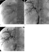
Pre-procedural antibiotic regimens, including a 3rd generation cephalosporin and metronidazole, were infused at least 1 hour prior to the procedure. Sedation was achieved with remifentanil, which was continuously infused by an infusion pump. The puncture was made with a 21G Skinny needle (PTC needle; A & A). If dilated peripheral ducts were visible on USG, a definitive puncture was performed with a 21G skinny needle under ultrasonographic guidance. If dilated peripheral ducts were not visible, a central duct was punctured under ultrasonographic guidance and the definitive puncture was made with another needle under fluoroscopic guidance. Following the definitive puncture, a 0.018 inch hairwire (A & A, Seongnam, Korea) was inserted through the needle to the bile duct. The hairwire was then changed to a 0.035-inch hydrophilic guide wire (Radifocus; Terumo, Tokyo, Japan) with the help of an introducing system (Yellow Sheath; A & A), which consisted of a 5F teflon dilator with an inner metal stiffener. The manipulation catheter was inserted over the 0.035-inch guide wire. After insertion of the manipulation catheter, the retained bile above the obstruction was aspirated, followed by cholangiographic evaluation with careful injection of contrast media. An attempt was made to cross all of the lesions, and the status of the bile duct beneath the obstructing lesion was also evaluated. For the hilar lesions, crossing the lesion was attempted to the contralateral duct and CBD. Finally, a drainage catheter was inserted into the most appropriate position to drain as much of the functioning liver as possible (Fig. 2).
Evaluation
The effectiveness of the catheter was evaluated based on the ability to aspirate retained bile and to cross the lesion. Crossing the lesion refers to passing the catheter into the duodenum in the CBD obstruction, and passing it into either the duodenum or contralateral lobe in the hilar obstruction. The safety of the catheter was evaluated based on whether or not insertion and manipulation of the catheter led to complications, and whether or not cholangitis was aggravated after the procedure. The severity of cholangitis was graded as follows: mild cholangitis was defined as laboratory testing, including a complete blood cell count, coagulation parameters, and renal function tests, within normal limits, as well as clear bile, whether or not it is black, yellow, or white in color; moderate cholangitis was defined as a fever >38° C with leukocytosis (>11,000>µL) despite initial medical treatment or turbid or purulent bile; and severe cholangitis was defined when there were signs of organ dysfunction.
RESULTS
Insertion of the manipulation catheter over a 0.035 hydrophilic guide wire was successful without a dilator in 85 of 91 patients (93.4%), of whom 82 underwent right access and 9 underwent left access. A 6F dilator was needed in 6 patients (6.6%; 5 for right access and 1 for left access). Aspiration of retained bile was successful in all patients (100%). The volume of aspirated bile ranged from 10-230 cc (mean, 59 cc). The aspirated bile was clear black (n=13), clear yellow (n=15), clear white (n=10), turbid green (n=25), or purulent (n=28).
The results of crossing obstructive lesions are summarized in Table 1. For the CBD obstructive lesions, crossing the lesion was successful in 98.1% of cases ( 1 case failed due to an impacted CBD stone). Crossing the lesion to the CBD in hilar obstructions was successful for 94.7% of cases (2 cases failed due to cholangiocarcinoma and iatrogenic surgical ligation). Crossing the lesion to the contralateral duct in hilar obstructions was successful in 92.1% of cases (3 cases failed due to cholangiocarcinoma).
Changes in the degree of cholangitis after PTBD are summarized in Table 2. Eighteen patients with mild cholangitis before the procedure improved within 3 days of the procedure. Of the 63 patients with moderate cholangitis, 2 patients (3%) had transient aggravation after PTBD, but improved within 7 days. Of the 10 patients with severe cholangitis, 9 (90%) improved within 7 days of PTBD and 1 (10%) improved 3 weeks after PTBD. In summary, cholangitis improved or in 89 of 91 patients (97%), and transiently aggravated in 2 patients (3%). Septic shock did not occur in any patients.
DISCUSSION
PTBD has been regarded as a drainage procedure for retained bile, rather than for patient evaluation, because of the potential to exacerbate cholangitis secondary to an elevation in intraductal pressure caused by catheter manipulation and contrast media. However, excessive restrictions for catheter manipulation in the biliary tract during the initial PTBD have limited the evaluation of obstructing lesions and bile duct anatomy, as well as directing the guide wire to the ideal position (12345678910). The most effective way to avoid elevation of intraductal pressure during PTBD is the aspiration of sufficient retained bile during the procedure (1234). However, the commonly used biliary introducing systems, which were designed to be inserted over a 0.018 inch wire are not suitable for bile aspiration, especially if there is blood-staining or thick purulent bile, because the introducing systems are thin and have an end-holed tip. Furthermore, these systems mostly consist of a straight dilator and an inner stiffener, so directional control is impossible. Another commercially available biliary manipulation catheter (Cook-Europe, Bjaeverskov, Denmark), which is a 6.5 Fr polyethylene catheter with a short angled tip, can facilitate directing of a wire with good torque control, but this is also unsuitable for aspiration of bile because it has a tapered end-holed tip (101112). Other angled tip angiographic catheters are helpful for directing a guide wire within the biliary tree, but the insertion of an angled angiographic catheter into the biliary tree can be difficult and even fail.
The biliary manipulation catheter used herein had sufficient stiffness and a tapered tip to allow for the easy insertion through a 0.035 inch guide wire. Only 6.6% of cases required a dilator for insertion of the catheter. The catheter was also effective in the aspiration of retained bile. We were able to aspirate nearly all of the retained bile with the catheter before further manipulation in all cases. The easily controllable tip and distal side holes improved the ability to aspirate, even through bile was purulent or blood-stained. Finally, good torque controllability was achieved with the short and stiff shaft, and the terminal angulated tip made it easy to cross lesions. For the CBD-obstructing lesions, we were able to cross all lesions except for one; due to a large impacted CBD stone. We did not try aggressive manipulation with the catheter in an attempt to avoid inducing pancreatitis. For the hilar-obstructing lesions, we were able to cross the lesion to the CBD in most cases (94.7%); the failed cases involved cholangiocarcinoma or an iatrogenic-ligated duct. On the second attempt, 2-3 days after the PTBD, we were successful in crossing all cholangiocarcinoma, but failed again with the ligated duct. The ligated duct was repaired by surgical revision. Crossing a wire to the contralateral duct is distinctly different from crossing a wire to the CBD, so we evaluated the contralateral crossing success rate separately. Contralateral crossing success is essential for either bilobar external drainage or bilobar T-configured stenting. We were also able to cross to the contralateral duct in most cases (92.1%), with the exception of 3 cholangiocarcimas, which had an acute (< 45 degrees) hilar angle. All the patients with a contralateral crossing failure were unresectable and underwent Y-configured bilobar stenting through the bilateral access.
There are a variety of different definitions of acute cholangitis in the literature. Some authors have defined acute cholangitis based on clinical signs, such as Charcot's triad (fever and/or chill, abdominal pain, and jaundice), while other authors have emphasized the properties of the bile. According to the Tokyo guidelines, the basic concepts of the criteria are as follows: 1) Charcot's triad is a definite diagnostic criterion for acute cholangitis; and 2) if a patient does not have all of the components of Charcot's triad, then a definite diagnosis can be achieved if both an inflammatory response and biliary obstruction are demonstrated by the laboratory data and imaging findings (13). Severity assessment criteria are based on the onset of organ dysfunction and response to initial medical treatment. Mild cholangitis is defined if there is response to initial medical treatment without organ dysfunction. Moderate cholangitis is defined if there is no response to initial medical treatment or organ dysfunction. Severe cholangitis is defined if there is organ dysfunction. Organs include cardiovascular, nervous, respiratory, renal, hepatic, and hematologic systems. As differentiation between mild and moderate cholangitis is ambiguous despite the above mentioned criteria, we made some modifications in the criteria based on blood test data and the properties of the aspirated bile during the procedure; specifically, if the body temperature was >38 ℃ with leukocytosis (>11,000>µL) despite initial medical treatment (including antibiotics) or the aspirated bile was turbid or purulent, severity was defined as moderate. In this study, cholangitis improved in 97% of patients, and was transiently aggravated in only 3% of patients. Septic shock did not occur in any patients.
Although the reported complication rates of PTBD are variable because of differences in techniques and reporting standards, hemobilia is known to occur in 2.6-9.0% of patients, and sepsis may occur in 5-26% of patients undergoing PTBD (12345678). In our study, minor bleeding occurred during needling in two patients, which was transient and controlled spontaneously. No bleeding complications occurred during insertion or manipulation of the catheter, as well as the following steps. With respect to cholangitic complications, our results were similar to or better than previous reports (12345678).
There were some limitations to this study. Precise classification of the severity of cholangitis was not possible during the follow-up period in some cases due to a lack of blood tests; such cases were categorized similar to the initial categorization. In addition, there was no control group which underwent PTBD without the use of a biliary manipulation catheter.
In conclusion, based on our results, the biliary manipulation catheter described herein is safe and effective for PTBD, attributed to the facilitation of bile aspiration and lesion crossing. The catheter can also be used for other percutaneous biliary procedures, such as percutaneous stone extraction and percutaneous stenting.


 XML Download